Your shopping cart is empty.
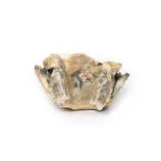
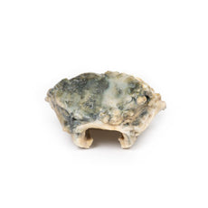
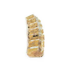
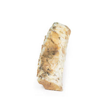
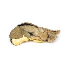
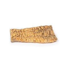
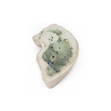
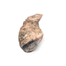
3D Printed Cerebral Arterio-Venous Malformation
Item # MP2010Need an estimate?
Click Add To Quote

-
by
A trusted GT partner -
3D Printed Model
from a real specimen -
Gov't pricing
Available upon request
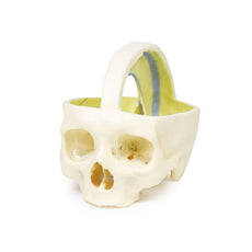
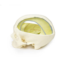
3D Printed Cerebral Arterio-Venous Malformation
Clinical History
This patient died at the age of 58 years from post-operative complications following
transurethral resection of the prostate. At the ages of 28 and 35, he had suffered two episodes of transient
neurological deficit. However, at 50 years, he developed permanent hemiparesis of the left leg chiefly affecting
his ankle.
Pathology
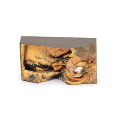
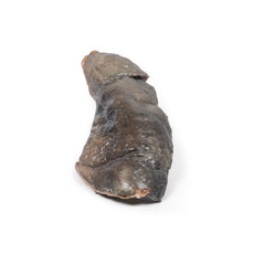
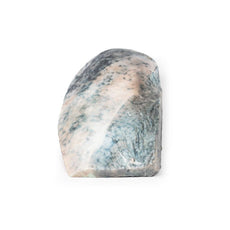
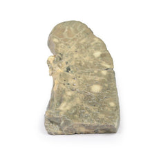
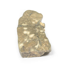
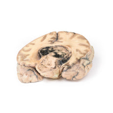
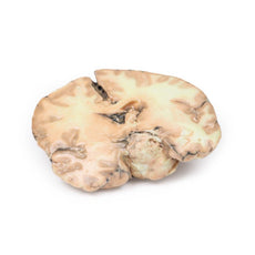
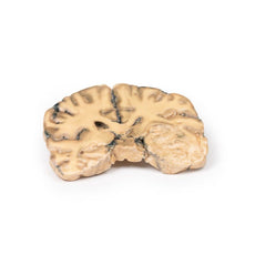
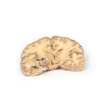
The specimen is a coronal slice of the brain that passes through the parietal lobes. Cortex and
white matter on the medial aspect of the right cerebral hemisphere have been replaced by a mass of abnormal
tissue 4 cm in greatest diameter. This lesion extends from the superior surface down to the roof of the lateral
ventricle. A closer inspection reveals the tissue to be a network of tortuous vascular channels and intervening
tissue.
Histological examination of this arterio-venous malformation showed glial tissue surrounding dilated
vessels. All vessels had a typical endothelial lining, some showed thick muscular walls and others thin walls,
thus identifying themselves as arteries and veins, respectively.
Further Information
The most frequently observed problems, related to cerebral arteriovenous malformations
(AVM), are headaches, seizures, cranial nerve deficits and back pain, and nausea may follow the occurrence of
coagulated blood escaping into the CSF in the vertebral column. Some patients with AVM have no symptoms at all.
Progressive weakness and numbness and vision changes as well as debilitating, excruciating pain may also occur
depending on the location of the AVMs. In serious cases, the vessels may rupture and cause intracranial
haemorrhage. In patients with AVM haemorrhage, symptoms caused by bleeding include loss of consciousness, sudden
and severe headache, nausea, vomiting, incontinence, and blurred vision, amongst others. Local damage on the
bleed site are also possible and can cause seizure, one-sided weakness (hemiparesis, as in this patient), a loss
of touch sensation on one side of the body, and deficits in language processing (aphasia). Ruptured AVMs are
responsible for considerable mortality and morbidity.
 Handling Guidelines for 3D Printed Models
GTSimulators by Global Technologies
Erler Zimmer Authorized Dealer
The models are very detailed and delicate. With normal production machines you cannot realize such details like shown in these models.
The printer used is a color-plastic printer. This is the most suitable printer for these models.
The plastic material is already the best and most suitable material for these prints. (The other option would be a kind of gypsum, but this is way more fragile. You even cannot get them out of the printer without breaking them).The huge advantage of the prints is that they are very realistic as the data is coming from real human specimen. Nothing is shaped or stylized.
The users have to handle these prints with utmost care. They are not made for touching or bending any thin nerves, arteries, vessels etc. The 3D printed models should sit on a table and just rotated at the table.


