Your shopping cart is empty.
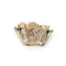
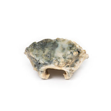
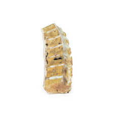
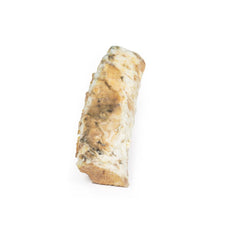
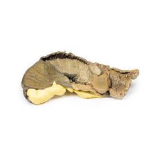
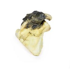
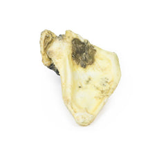
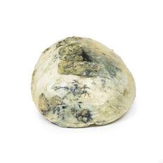
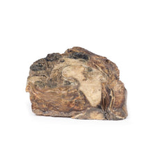
3D Printed Pedunculated Adenoma of the Colon
Item # MP2081Need an estimate?
Click Add To Quote

-
by
A trusted GT partner -
3D Printed Model
from a real specimen -
Gov't pricing
Available upon request
3D Printed Pedunculated Adenoma of the Colon
Clinical History
A 50-year old male underwent a colonoscopy after testing positive for faecal
occult blood during a screening test. Colonoscopy revealed a pedunculated tumour in the descending colon, which was
later resected.
Pathology
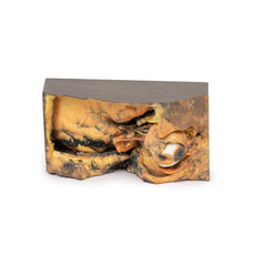
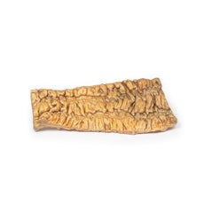
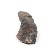
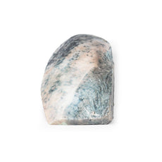
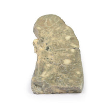
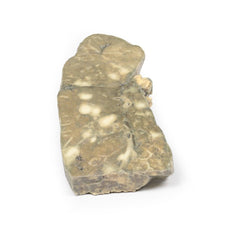
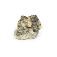
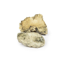
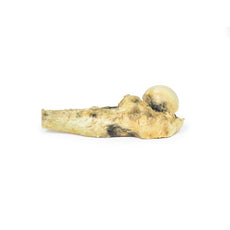
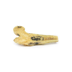
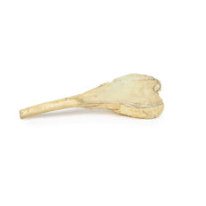
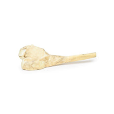
This specimen is the resected segment of descending colon. There is a single dark
lobulated mass visible arising from the mucosal surface. It is attached to a stalk which is 4cm in length.
Histologically, the mass comprises a core of connective tissue covered with hyperplastic glandular epithelium of
colonic type, with focal nuclear atypia. This is an example of a tubular colonic adenoma.
Further Information
Colorectal adenomas are intraepithelial neoplasms that characteristically
display epithelial dysplasia. They are benign but are precursors to adenocarcinoma. Not all adenomas evolve into
adenocarcinoma. They produce polyps (sometimes pedunculated) or sessile lesions or variable size. They occur
predominantly in males and are more common in Western countries due to diet and lifestyle. They are present in about
30% of people over the age of 60 years in the West. There is an increased risk in patients with a positive family
history of colorectal adenocarcinoma. Regular surveillance colonoscopy in at risk groups with polyp removal reduces
incidence of adenocarcinoma. There are three classifications of colonic adenomas based on their architecture:
tubular (>75% have a tubular morphology), tubulovillous (25-75% villous morphology) and villous (>75% have
villous morphology). Histologically, they may have epithelial dysplasia characterized by nuclear hyperchromasia,
elongation and stratification. Tubular adenomas tend to be small, pedunculated polys composed of rounded or tubular
glands. Pedunculated adenomas have a slender fibromuscular stalk with blood vessels derived from the submucosa. The
stalk is usually non-neoplastic epithelium. The size of the adenoma is the biggest predictor of progression to
adenocarcinoma. Progression is rare in adenomas <1cm in diameter. However, up to 40% of lesions larger than 4cm
in diameter progress to adenocarcinoma.
Most adenomas are asymptomatic and slow growing. Large polyps may present with symptoms of anaemia from occult
bleeding. Villous adenomas occasionally secrete large amounts of mucoid protein and/or potassium rich fluid, leading
possibly to hypokalemia.
 Handling Guidelines for 3D Printed Models
GTSimulators by Global Technologies
Erler Zimmer Authorized Dealer
The models are very detailed and delicate. With normal production machines you cannot realize such details like shown in these models.
The printer used is a color-plastic printer. This is the most suitable printer for these models.
The plastic material is already the best and most suitable material for these prints. (The other option would be a kind of gypsum, but this is way more fragile. You even cannot get them out of the printer without breaking them).The huge advantage of the prints is that they are very realistic as the data is coming from real human specimen. Nothing is shaped or stylized.
The users have to handle these prints with utmost care. They are not made for touching or bending any thin nerves, arteries, vessels etc. The 3D printed models should sit on a table and just rotated at the table.


