Your shopping cart is empty.
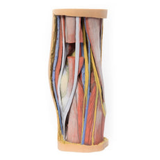
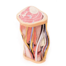
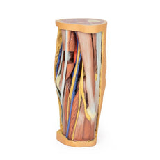
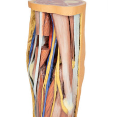
3D Printed Arm, Forearm and Hand Replica
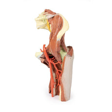
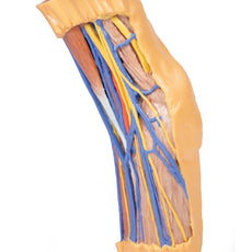
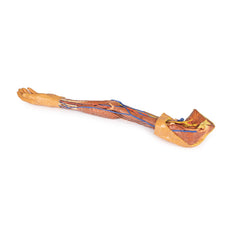
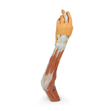
In the distal arm and elbow/cubital fossa region we can see the arrangement of the biceps tendon, brachial artery and median nerve from lateral to medial. The bicipital aponeurosis has been divided to reveal the structures deep to it. The ulnar nerve can be seen passing behind the medial epicondy
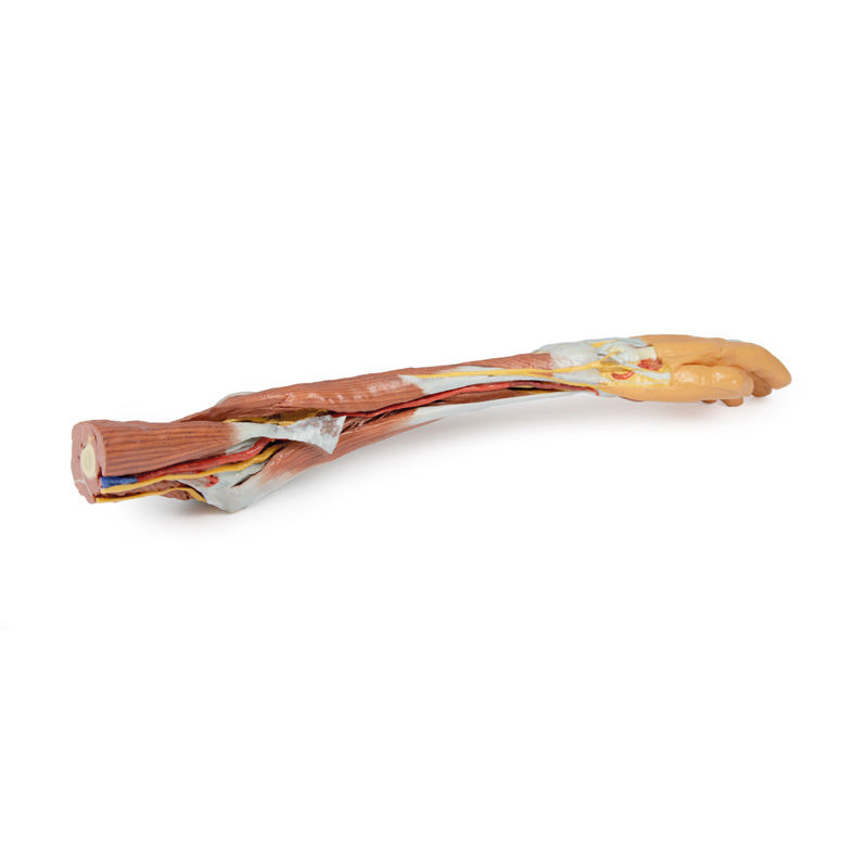
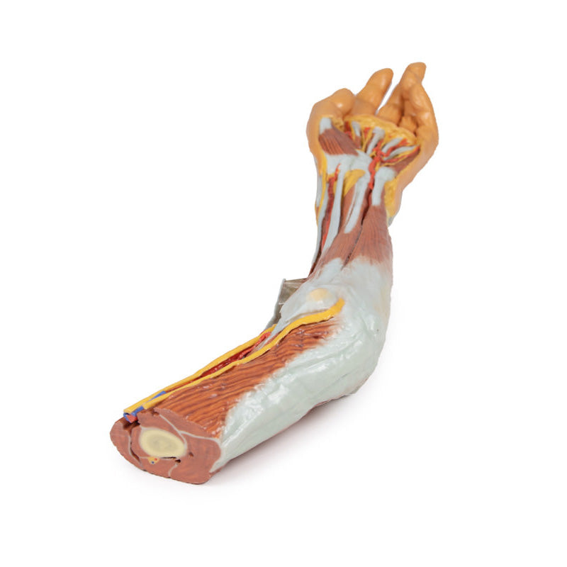
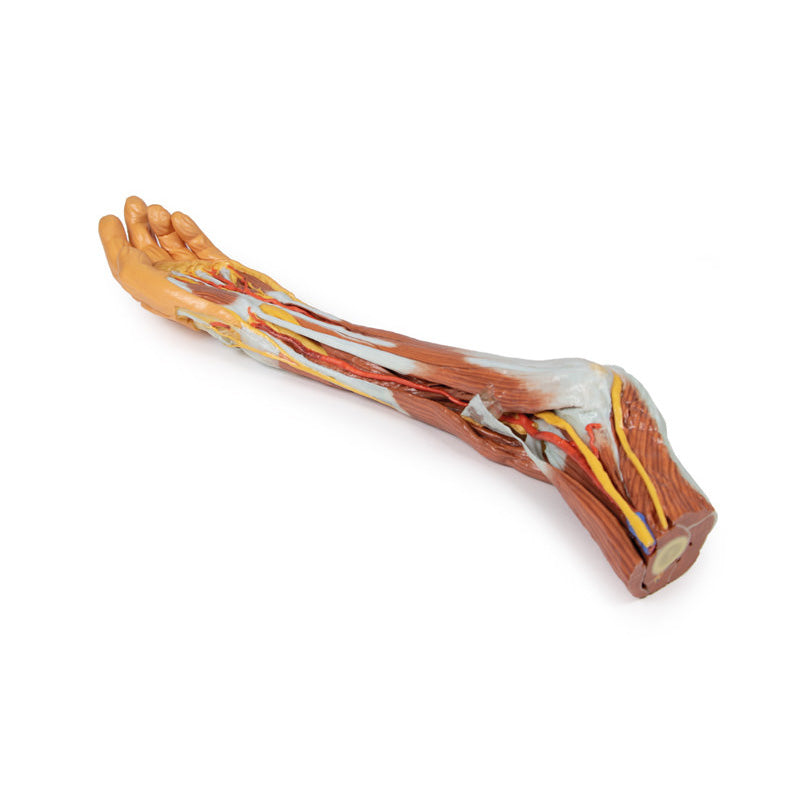
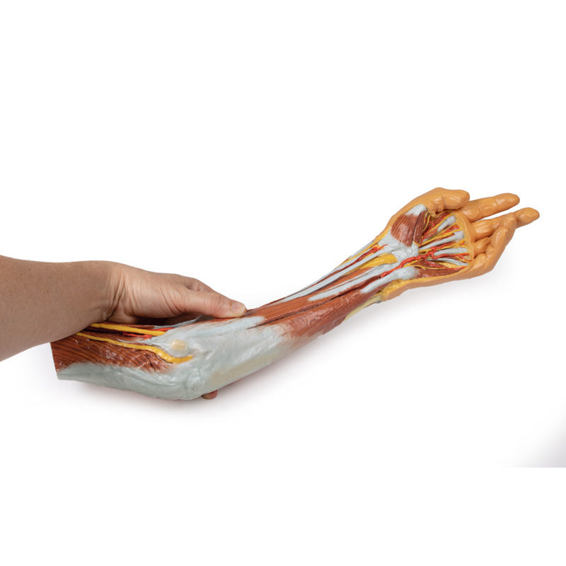
In the distal arm and elbow/cubital fossa region we can see the arrangement of the biceps tendon, brachial artery and median nerve from lateral to medial. The bicipital aponeurosis has been divided to reveal the structures deep to it. The ulnar nerve can be seen passing behind the medial epicondyle with an ulnar collateral artery close by. The superficial branch of the radial nerve can just be seen in the space between brachioradialis and brachialis muscles (as the belly of the latter muscle has been displaced slightly laterally).
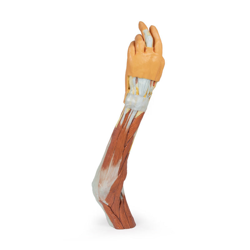
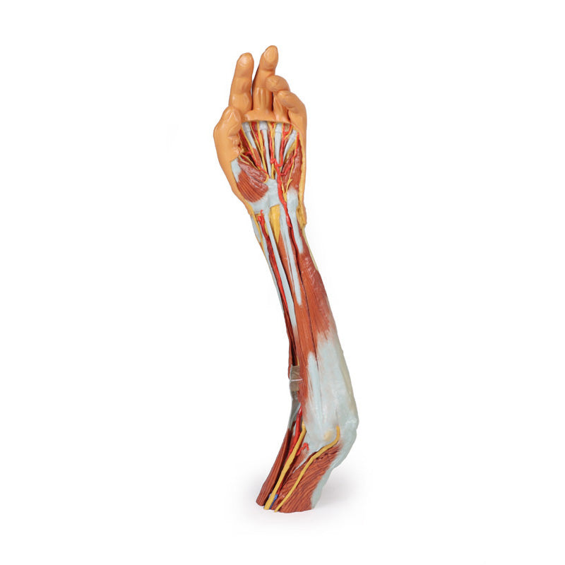
In the forearm, the superficial flexor muscles arising from the common flexor origin can be clearly seen (from lateral to medial– pronator teres, flexor carpi radialis (FCR), flexor digitorum superficialis (FDS) and flexor carpi ulnaris (FCU)). There is not a palmaris longus muscle in this cadaver. The radial artery and superficial branch of the radial nerve (emerging half way down the forearm from behind the brachioradialis muscle and tendon) are clearly identifiable. The ulnar artery can be seen in the distal forearm emerging from beneath FCU muscle.
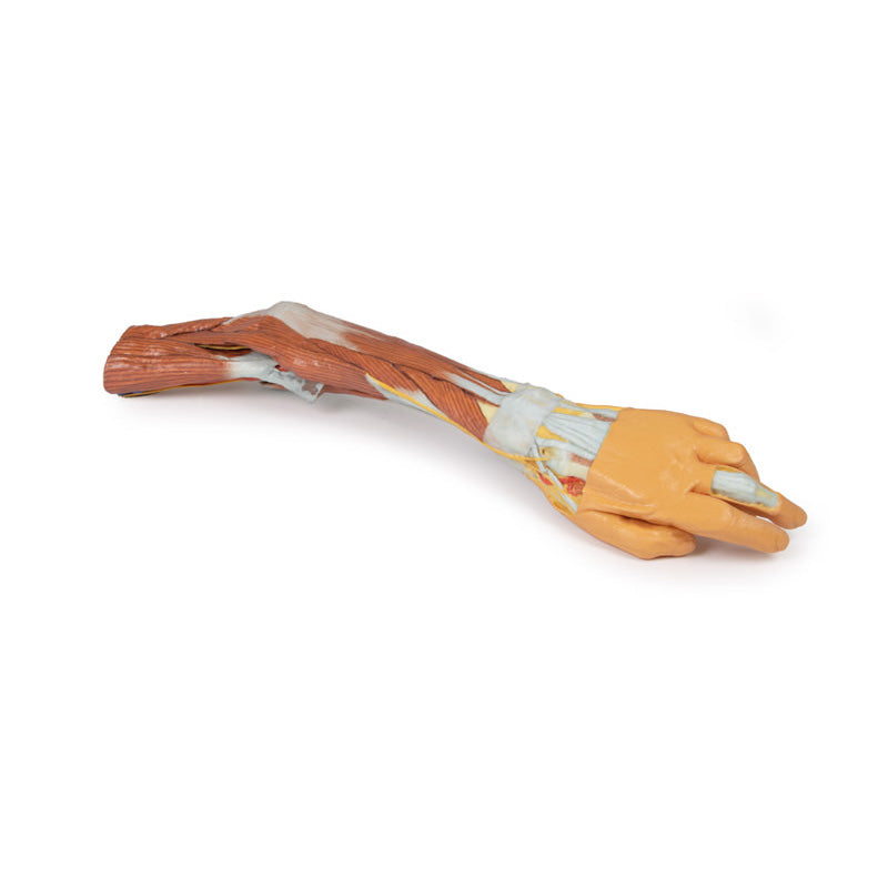
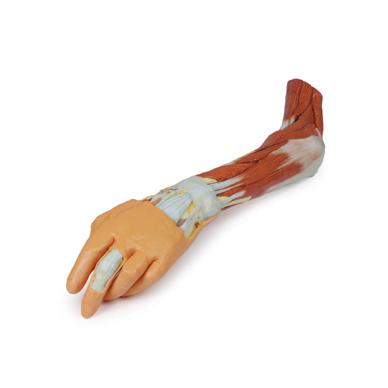
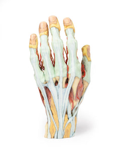
On the posterior aspect of the forearm the extensor muscles arising from the common extensor origin are clearly identifiable. These include (from medial to lateral) the extensor carpi ulnaris (ECU), extensor digiti minimi, extensor digitorum and extensor carpi radialis brevis (ECRB). The extensor carpi radialis longus (ECRL) can be seen arising from the inferior aspect of the lateral supracondylar ridge. Further distally the abductor pollicis longus (APL) and extensor pollicis brevis (EPB) can be seen emerging from deep to superficial and ‘wrapping’ around the radius. They along with extensor pollicis longus (EPL) (partly hidden) travel distally to insert into the extensor or dorsal surface of the base of the 1st metacarpal, proximal phalanx and distal phalanx of the thumb, respectively. The anatomical snuffbox is displayed with the radial artery in its floor (surrounded by fat) and the cutaneous branch of the radial nerve in its roof. The extensor retinaculum is clearly visible on the dorsum of the wrist and distal to it the tendons of extensor indicis and ECRB and ECRL can be seen inserting into the 2nd and 3rd metacarpals.
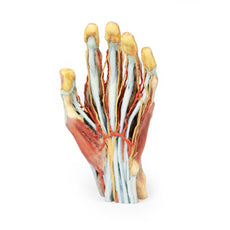
In the hand, the superficial dissection reveals muscles of the thenar and hypothenar eminences, the flexor retinaculum of the hand (roof of the carpal tunnel), the long tendons of the hand, the lumbricals, and the superficial palmar arch arising from the ulnar artery, which passes into the hand lateral to the pisiform bone above the retinaculum, along with the superficial branch of the ulnar nerve. The large median nerve can be seen passing beneath the flexor retinaculum between the FCR and the FDS tendons. Digital arteries and nerves can be clearly seen further distally in the palm entering the digits. Note in particular the small recurrent branch of the median nerve crossing over the flexor pollicis brevis close to its origin from the retinaculum. The extensor expansion is dissected on the middle finger.
Download Handling Guidelines for 3D Printed Models
GTSimulators by Global Technologies
Erler Zimmer Authorized Dealer

9.0 lb
3D Printed Arm, Forearm and Hand Replica
Item # MP1510
$1,704.00
$1,894.00
You save $190.00
Need an estimate?
Click Add To Quote

Features & Specifications
-
by
A trusted GT partner -
FREE Shipping
U.S. Contiguous States Only -
3D Printed Model
from a real specimen -
Gov't pricing
Available upon request
About This Item
3D Printed Arm, Forearm and Hand Replica
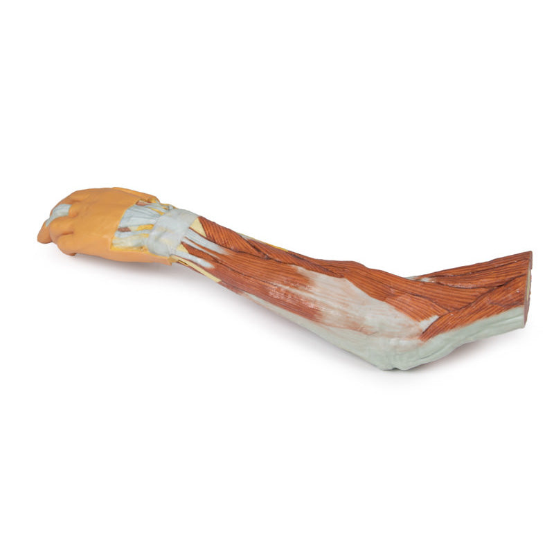
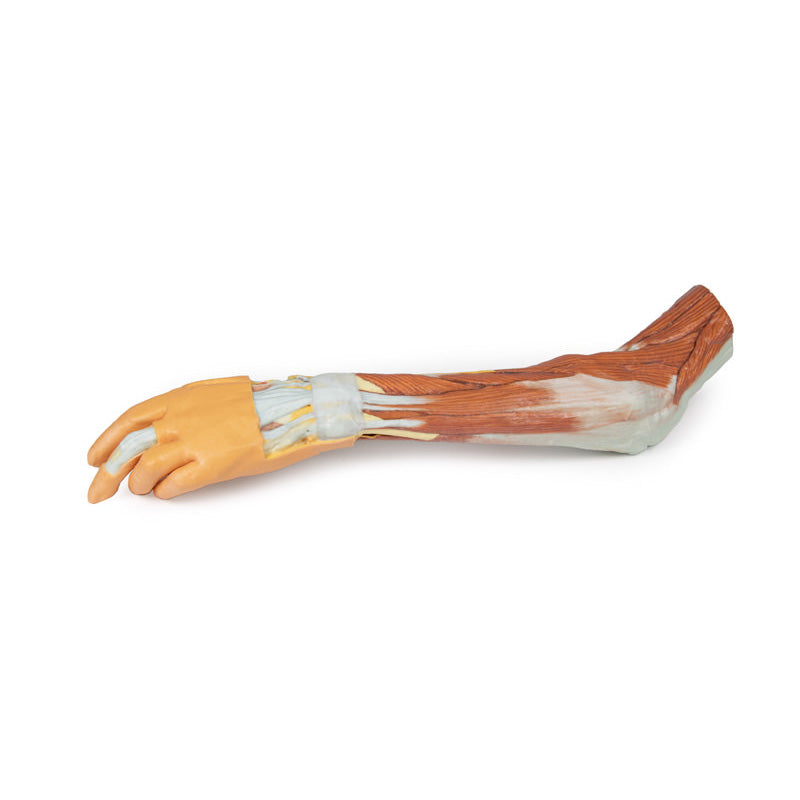
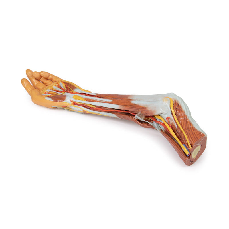
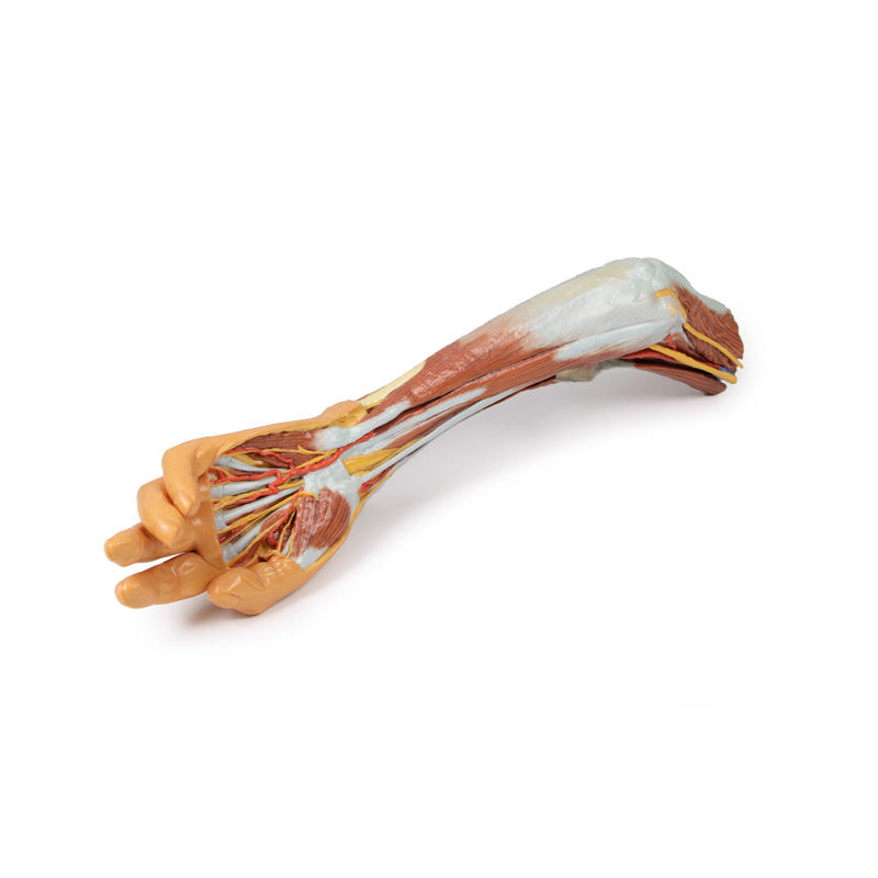
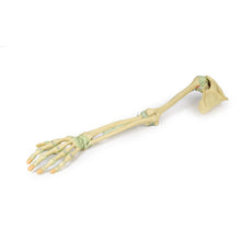
This upper limb specimen displays the vascular, nervous and muscular anatomy of a left distal arm, forearm and hand.In the distal arm and elbow/cubital fossa region we can see the arrangement of the biceps tendon, brachial artery and median nerve from lateral to medial. The bicipital aponeurosis has been divided to reveal the structures deep to it. The ulnar nerve can be seen passing behind the medial epicondy
3D Printed Arm, Forearm and Hand Replica
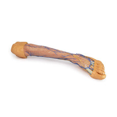
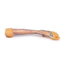
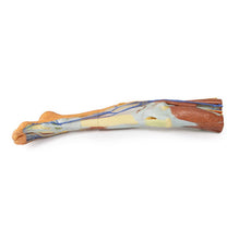
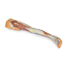
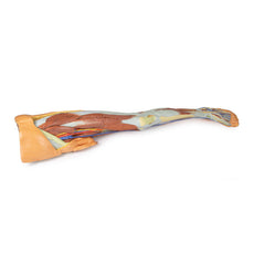
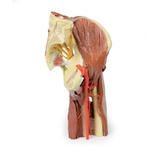
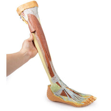
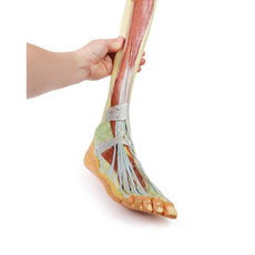
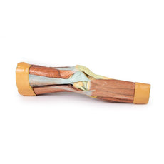
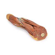
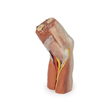
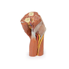
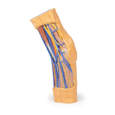
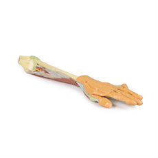
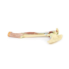
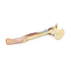
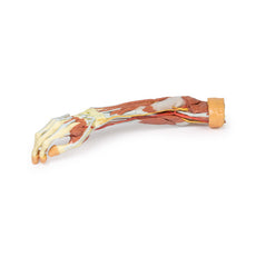
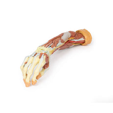
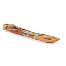
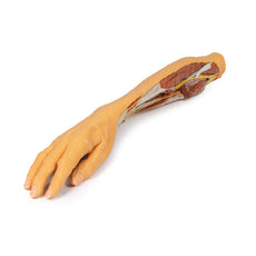
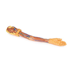
This upper limb specimen displays the vascular, nervous and muscular anatomy of a left distal arm, forearm and hand.In the distal arm and elbow/cubital fossa region we can see the arrangement of the biceps tendon, brachial artery and median nerve from lateral to medial. The bicipital aponeurosis has been divided to reveal the structures deep to it. The ulnar nerve can be seen passing behind the medial epicondyle with an ulnar collateral artery close by. The superficial branch of the radial nerve can just be seen in the space between brachioradialis and brachialis muscles (as the belly of the latter muscle has been displaced slightly laterally).
In the forearm, the superficial flexor muscles arising from the common flexor origin can be clearly seen (from lateral to medial– pronator teres, flexor carpi radialis (FCR), flexor digitorum superficialis (FDS) and flexor carpi ulnaris (FCU)). There is not a palmaris longus muscle in this cadaver. The radial artery and superficial branch of the radial nerve (emerging half way down the forearm from behind the brachioradialis muscle and tendon) are clearly identifiable. The ulnar artery can be seen in the distal forearm emerging from beneath FCU muscle.
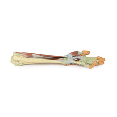
On the posterior aspect of the forearm the extensor muscles arising from the common extensor origin are clearly identifiable. These include (from medial to lateral) the extensor carpi ulnaris (ECU), extensor digiti minimi, extensor digitorum and extensor carpi radialis brevis (ECRB). The extensor carpi radialis longus (ECRL) can be seen arising from the inferior aspect of the lateral supracondylar ridge. Further distally the abductor pollicis longus (APL) and extensor pollicis brevis (EPB) can be seen emerging from deep to superficial and ‘wrapping’ around the radius. They along with extensor pollicis longus (EPL) (partly hidden) travel distally to insert into the extensor or dorsal surface of the base of the 1st metacarpal, proximal phalanx and distal phalanx of the thumb, respectively. The anatomical snuffbox is displayed with the radial artery in its floor (surrounded by fat) and the cutaneous branch of the radial nerve in its roof. The extensor retinaculum is clearly visible on the dorsum of the wrist and distal to it the tendons of extensor indicis and ECRB and ECRL can be seen inserting into the 2nd and 3rd metacarpals.
In the hand, the superficial dissection reveals muscles of the thenar and hypothenar eminences, the flexor retinaculum of the hand (roof of the carpal tunnel), the long tendons of the hand, the lumbricals, and the superficial palmar arch arising from the ulnar artery, which passes into the hand lateral to the pisiform bone above the retinaculum, along with the superficial branch of the ulnar nerve. The large median nerve can be seen passing beneath the flexor retinaculum between the FCR and the FDS tendons. Digital arteries and nerves can be clearly seen further distally in the palm entering the digits. Note in particular the small recurrent branch of the median nerve crossing over the flexor pollicis brevis close to its origin from the retinaculum. The extensor expansion is dissected on the middle finger.
Download Handling Guidelines for 3D Printed Models
GTSimulators by Global Technologies
Erler Zimmer Authorized Dealer
These items normal warranty are two years, however the warranty doesn’t cover “wear and tear”. The manufacturer does have 100% quality control on these models.
The models are very detailed and delicate. With normal production machines you cannot realize such details like shown in these models.
The printer used is a color-plastic printer. This is the most suitable printer for these models.
The plastic material is already the best and most suitable material for these prints. (The other option would be a kind of gypsum, but this is way more fragile. You even cannot get them out of the printer without breaking them).The huge advantage of the prints is that they are very realistic as the data is coming from real human specimen. Nothing is shaped or stylized.
The users have to handle these prints with utmost care. They are not made for touching or bending any thin nerves, arteries, vessels etc. The 3D printed models should sit on a table and just rotated at the table.

by — Item # MP1510
3D Printed Arm, Forearm and Hand Replica
$1,704.00
$1,894.00
Add to Cart
Add to Quote


