Your shopping cart is empty.
3D Printed Male Pelvis
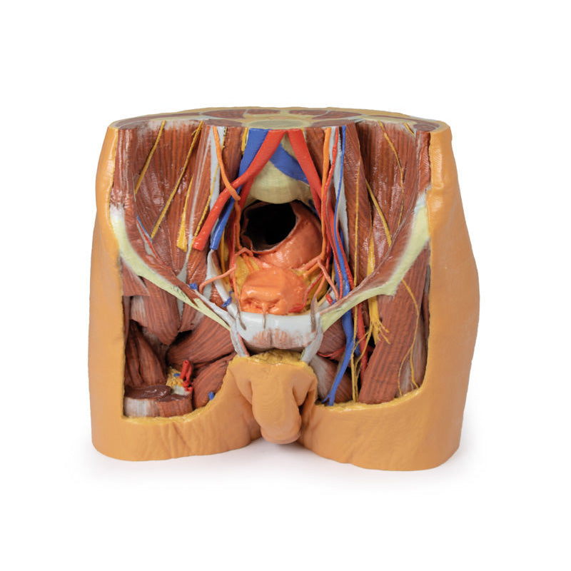
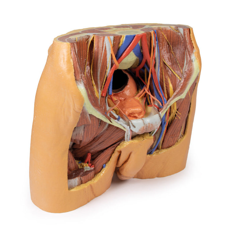
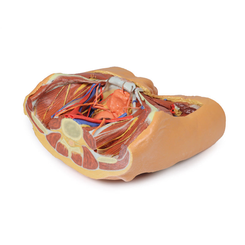
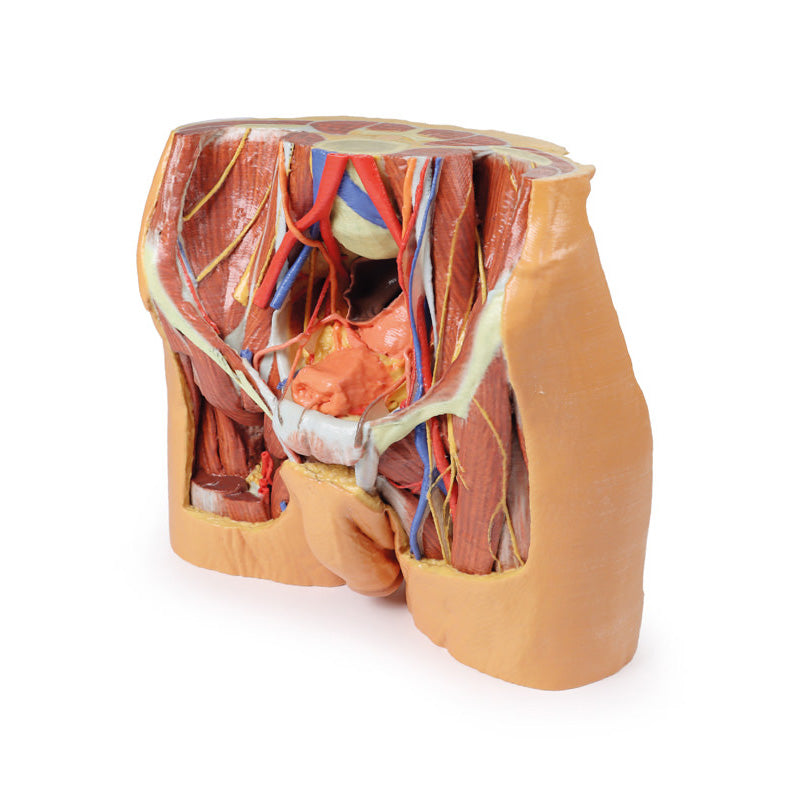
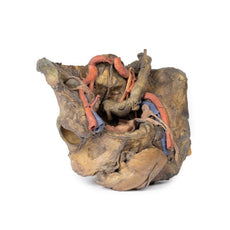
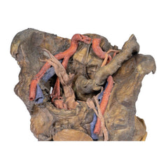
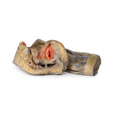
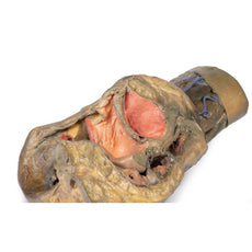
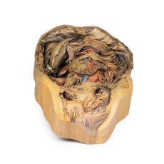
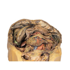
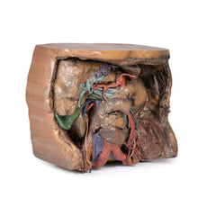
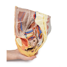
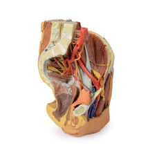
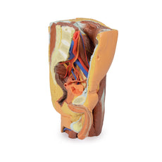
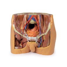
Lower posterior abdominal wall and false pelvis: The specimen is transected at approximately the level of the L2/L3 intervertebral disc. The common iliac veins unite to form the inferior vena cava. The common iliac arteries are close to uniting at the top of the print. The iliacus and psoas muscles are easy to identify, the latter has a prominent psoas minor tendon. They can be seen to unite as they pass under the inguinal ligament. The nerves of the iliac fossa (from superior to inferior: ilioinguinal nerve, lateral cutaneous nerve of thigh, femoral nerve ) and their course is clearly visible, as is the genitofemoral nerves on the surface of psoas muscle. The ureters also descend on the superficial surface of the psoas and cross from its lateral to its medial border. They enter the pelvis at the bifurcation of the common iliac arteries into external and internal arteries. The external iliac arteries and veins running along the pelvic brim are clearly visible, as is the vas deferens crossing the brim from the deep inguinal ring to enter the pelvis.
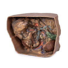
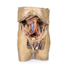
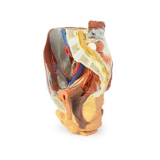
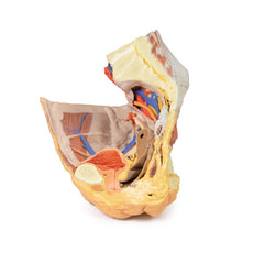
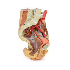
True pelvis: The pelvis is dominated by a dilated rectum, dissected to demonstrate a transverse fold. The bladder is seen anteriorly in the pelvis (with the obliterated urachus passing towards the anterior abdominal wall) and the ureters can be seen entering the bladder wall posteriorly. The branches of the internal iliac artery can be clearly seen, with the obturator exiting the pelvis through the obturator foramen with its accompanying artery and vein. There is an accessory obturator vein crossing the brim in addition to the usual branch which drains to the internal iliac vein. The obliterated umbilical arteries are seen exiting the pelvis anteriorly and ascending on the anterior abdominal wall (reflected anteriorly).
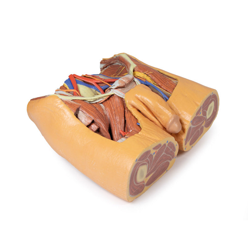
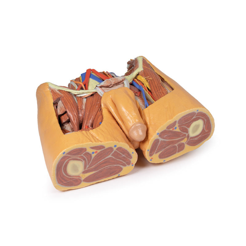
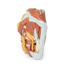
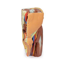
The femoral triangle: On the right the muscles on the floor of the triangle are dissected. On the left the vein, artery and nerve have been retained as they pass deep to the inguinal ligament.
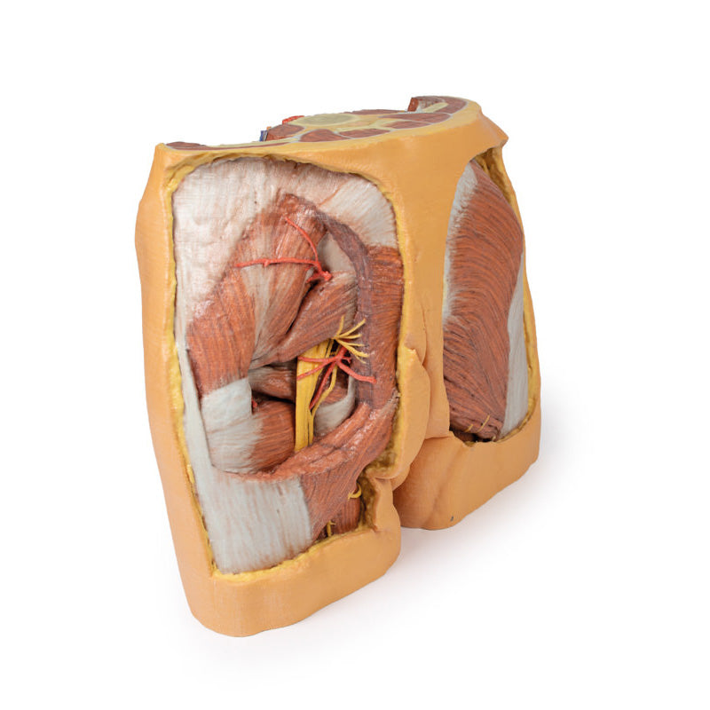
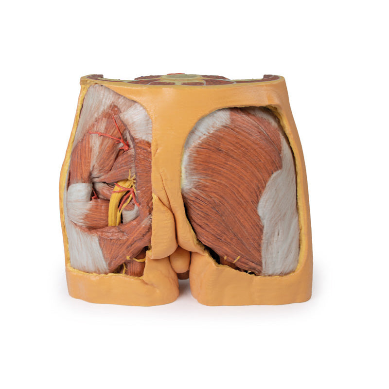
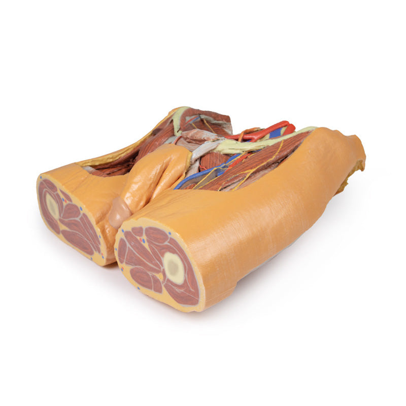
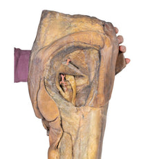
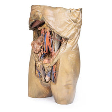
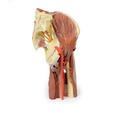
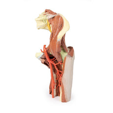
Gluteal region: The right gluteal region is dissected down to the gluteus maximus and no further. The perforating cutaneous nerves (S2-S3)/cutaneous branches of the inferior gluteal nerve can be seen winding around the lower edge of the gluteus maximus muscle. The extensive origin of the gluteus maximus is readily seen and its course inferiorly to its insertion on the femur is visible (though not the actual insertion). The tensor fascia lata and iliotibial tract are evident on the lateral aspect. On the left a ‘window’ has been made in the gluteus maximus to reveal the deeper lying gluteus medius and piriformis. The sciatic nerve arises deep to piriformis, and passes superficially to the superior and inferior gemelli, obturator internus and the quadratus femoris muscles. Descending adjacent to the sciatic nerve is the inferior gluteal nerve with its accompanying artery. The inferior cluneal nerve and perineal branch of the posterior cutaneous nerve of thigh can be seen just briefly lying above semitendinosus. The superior gluteal artery can be seen just superior to the piriformis.
Download Handling Guidelines for 3D Printed Models
GTSimulators by Global Technologies
Erler Zimmer Authorized Dealer

13.0 lb
3D Printed Male Pelvis
Item # MP1770
$6,290.00
$6,989.00
You save $699.00
Need an estimate?
Click Add To Quote

Features & Specifications
-
by
A trusted GT partner -
FREE Shipping
U.S. Contiguous States Only -
3D Printed Model
from a real specimen -
Gov't pricing
Available upon request
Frequently Bought Together
3D Printed Male Pelvis
This multipart 3D printed specimen represents the inferior portions of our larger posterior abdominal wall print (MP1300) that displays the inferior posterior abdominal wall, the pelvic cavity and the proximal thigh (including the gluteal regions and femoral triangles).Lower posterior abdominal wall and false pelvis: The specimen is transected at approximately the level of the L2/L3 intervertebral disc. The common iliac veins unite to form the inferior vena cava. The common iliac arteries are close to uniting at the top of the print. The iliacus and psoas muscles are easy to identify, the latter has a prominent psoas minor tendon. They can be seen to unite as they pass under the inguinal ligament. The nerves of the iliac fossa (from superior to inferior: ilioinguinal nerve, lateral cutaneous nerve of thigh, femoral nerve ) and their course is clearly visible, as is the genitofemoral nerves on the surface of psoas muscle. The ureters also descend on the superficial surface of the psoas and cross from its lateral to its medial border. They enter the pelvis at the bifurcation of the common iliac arteries into external and internal arteries. The external iliac arteries and veins running along the pelvic brim are clearly visible, as is the vas deferens crossing the brim from the deep inguinal ring to enter the pelvis.
True pelvis: The pelvis is dominated by a dilated rectum, dissected to demonstrate a transverse fold. The bladder is seen anteriorly in the pelvis (with the obliterated urachus passing towards the anterior abdominal wall) and the ureters can be seen entering the bladder wall posteriorly. The branches of the internal iliac artery can be clearly seen, with the obturator exiting the pelvis through the obturator foramen with its accompanying artery and vein. There is an accessory obturator vein crossing the brim in addition to the usual branch which drains to the internal iliac vein. The obliterated umbilical arteries are seen exiting the pelvis anteriorly and ascending on the anterior abdominal wall (reflected anteriorly).
The femoral triangle: On the right the muscles on the floor of the triangle are dissected. On the left the vein, artery and nerve have been retained as they pass deep to the inguinal ligament.
Gluteal region: The right gluteal region is dissected down to the gluteus maximus and no further. The perforating cutaneous nerves (S2-S3)/cutaneous branches of the inferior gluteal nerve can be seen winding around the lower edge of the gluteus maximus muscle. The extensive origin of the gluteus maximus is readily seen and its course inferiorly to its insertion on the femur is visible (though not the actual insertion). The tensor fascia lata and iliotibial tract are evident on the lateral aspect. On the left a ‘window’ has been made in the gluteus maximus to reveal the deeper lying gluteus medius and piriformis. The sciatic nerve arises deep to piriformis, and passes superficially to the superior and inferior gemelli, obturator internus and the quadratus femoris muscles. Descending adjacent to the sciatic nerve is the inferior gluteal nerve with its accompanying artery. The inferior cluneal nerve and perineal branch of the posterior cutaneous nerve of thigh can be seen just briefly lying above semitendinosus. The superior gluteal artery can be seen just superior to the piriformis.
Download Handling Guidelines for 3D Printed Models
GTSimulators by Global Technologies
Erler Zimmer Authorized Dealer
These items normal warranty are two years, however the warranty doesn’t cover “wear and tear”. The manufacturer does have 100% quality control on these models.
The models are very detailed and delicate. With normal production machines you cannot realize such details like shown in these models.
The printer used is a color-plastic printer. This is the most suitable printer for these models.
The plastic material is already the best and most suitable material for these prints. (The other option would be a kind of gypsum, but this is way more fragile. You even cannot get them out of the printer without breaking them).The huge advantage of the prints is that they are very realistic as the data is coming from real human specimen. Nothing is shaped or stylized.
The users have to handle these prints with utmost care. They are not made for touching or bending any thin nerves, arteries, vessels etc. The 3D printed models should sit on a table and just rotated at the table.

by — Item # MP1770
3D Printed Male Pelvis
$6,290.00
$6,989.00
Add to Cart
Add to Quote


