Your shopping cart is empty.
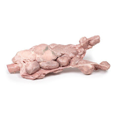
3D Printed Aorta & Para-aortic Lymph Nodes
Item # MP2103Need an estimate?
Click Add To Quote

-
by
A trusted GT partner -
FREE Shipping
U.S. Contiguous States Only -
3D Printed Model
from a real specimen -
Gov't pricing
Available upon request
3D Printed Aorta & Para-aortic Lymph Nodes
Clinical History
This was a case of a 75-year-old female who presented with symptoms of
recurrent disease and confirmed to have chemo-resistant multiple retroperitoneal lymph node metastases five years
after the initial therapy for stage IIIc serous adenocarcinoma of the ovary. Positron emission tomography/computed
tomography (PET/CT) revealed the involvement of para-aortic nodes and pelvic nodes. She died of liver complications
before therapeutic options, such as radical lymphadenectomy, could be considered.
Pathology
The specimen consists of the abdominal aorta and common iliac arteries surrounded by
large numbers of extremely enlarged iliac nodes para-aortic lymph nodes. Histopathological examination revealed
metastatic high-grade adenocarcinoma in some of the resected lymph nodes.
Further information
Occasionally, lymph node metastases represent the only component at the time
of recurrence of ovarian cancer. In this case, the metastatic nodes predicted by PET/CT completely corresponded to
the actual metastatic nodes. Ultrasound (US) could equally have confirmed the presence of such large lymph nodes.
PET/CT or US often fails to identify microscopic disease in histopathologically-proven positive nodes. Therefore, it
is difficult to reliably exclude lymph node metastases during surveillance following initial surgery for ovarian
cancer.
In the context of a recurrent ovarian disease, systematic aortic and pelvic node dissection would be considered
appropriate in younger women with no other evidence of metastatic disease. This is unlikely to be curative but may
produce palliation with symptom control and allow for the trial of novel therapies if available.
More commonly,
sampling of pelvic and aortic lymph nodes is part of the formal surgical staging of epithelial ovarian cancer at the
time of initial surgical treatment. In addition, in women presenting initially with advanced stage ovarian cancer,
systematic debulking of enlarged retroperitoneal lymph nodes will be considered if this leads to complete debulking
of the tumour.
 Handling Guidelines for 3D Printed Models
GTSimulators by Global Technologies
Erler Zimmer Authorized Dealer
The models are very detailed and delicate. With normal production machines you cannot realize such details like shown in these models.
The printer used is a color-plastic printer. This is the most suitable printer for these models.
The plastic material is already the best and most suitable material for these prints. (The other option would be a kind of gypsum, but this is way more fragile. You even cannot get them out of the printer without breaking them).The huge advantage of the prints is that they are very realistic as the data is coming from real human specimen. Nothing is shaped or stylized.
The users have to handle these prints with utmost care. They are not made for touching or bending any thin nerves, arteries, vessels etc. The 3D printed models should sit on a table and just rotated at the table.


