Your shopping cart is empty.
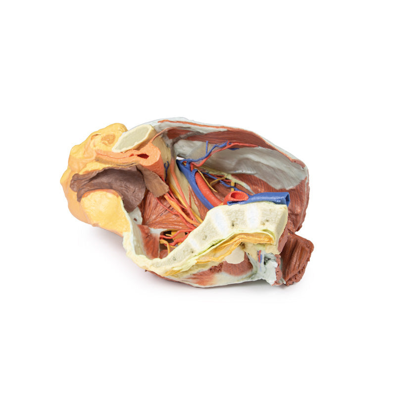
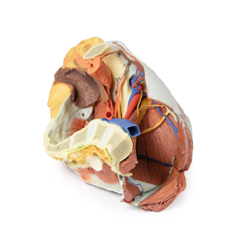
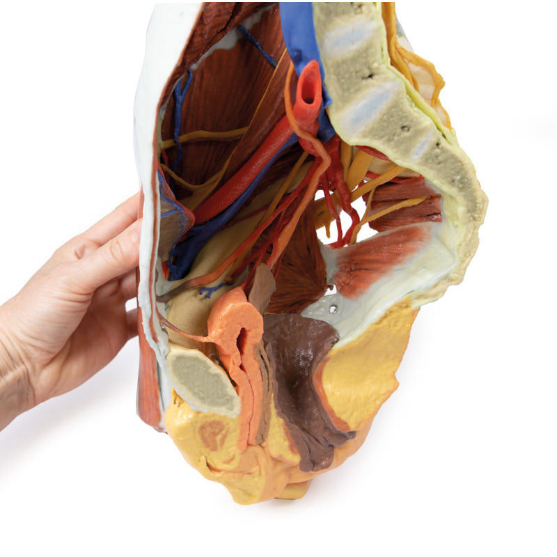
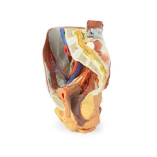
3D Printed Female right pelvis
Internal anatomy:
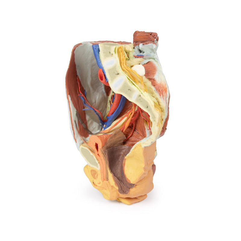
The muscular boundaries of the inferior abdominal cavity are defined posterolaterally bythe quadratus lumborum, iliacus and psoas muscles; anteriorly by the (varyinglyexposed) external and internal abdominal oblique muscles, the transversus abdominis andrectus abdominis. Inferiorly in the pelvic cavity, the obturator internus is visible traversingthrough the lesser sciatic foramen inferior to the sacrospinous ligament. Fibres of coccygeusmerge with those of the sacrospinous ligament. Piriformis has been sectioned, with bothorigin visible within the cavity (and part visible in the gluteal region). The common iliac artery arises from its cut edge at the level of L5, bifurcating at the level ofthe sacral promontory into the external and internal iliac arteries.
The external iliac arterycrosses the pelvic brim to give off the deep circumflex iliac artery and inferior epigastricartery before exiting the pelvis deep to the inguinal ligament. The internal iliac artery runsposterolaterally, giving the iliolumbar artery posteriorly and lateral sacral arteries whichenter the anterior sacral foramina.
A radicular artery entering the anterior foramina of thecoccyx can also be seen. Inferiorly, the superior gluteal artery, inferior gluteal artery andinternal pudendal artery exit the pelvic cavity through the greater sciatic foramen. A branchfrom the inferior gluteal artery, supplying psoas, travels anteriorly along the pectineal line.Anteriorly, the umbilical artery gives off the superior vesical artery (supplying the bladder)before terminating against the anterior abdominal wall as the medial umbilical ligament. Posteriorly, the inferior vesical artery arises from the obturator artery beforeexiting the pelvis through the obturator canal. The uterine artery crosses over the ureter toenter the remnants of the broad ligament.
The major veins preserved are the inferior epigastric vein and deep circumflex iliac veindraining into the external iliac vein, and the iliolumbar vein and lateral sacral vein draininginto the internal iliac vein. The external iliac vein and internal iliac vein unite to form theright common iliac vein which, at the level of L5, joins the (cut edge) of the left common iliacvein to become the inferior vena cava. Two veins pass horizontally across iliacus andquadratus lumborum.
Of the peripheral nerve structures preserved in this specimen, the lateral femoral cutaneousnerve passes laterally across the superficial aspect of the iliacus muscle and the femoralnerve is visible deep to the psoas major muscle. The genitofemoral nerve lies on thesuperficial surface of the psoas major, and the course of the genital branch entering thedeep inguinal ring and the femoral branch passing deep to the inguinal ligament can befollowed. The obturator nerve is also seen passing from deep to the psoas muscle anteriorlyto the obturator foramen. In the true pelvis the lumbosacral trunk crosses the pelvic brimand joints the anterior ramus of S1.
The S1-S3 anterior rami are visible and can be followedas they pass through the greater sciatic foramen and enter the gluteal region. In addition to the muscular and neurovascular structures, parts of pelvic viscera have beenpreserved. Posterior to the pubic symphysis the median umbilical ligament extendssuperiorly from the bladder to the anterior abdominal wall. The ureter descends anteriorlyto the psoas major muscle, across the iliac vessels, and beneath the uterine artery to enterthe posterior bladder wall. The urethra is seen passing downwards to its opening at theurinary meatus, just posterior to the clitoris. Posterior to the bladder are the posthysterectomyremnants of the uterus at the apex of the closed, superior end of the vagina.Posterior to this part of the cut rectum, anal canal and anus are present. Some muscularfibres of levator ani and the external anal sphincter can be seen in the ischioanal fossa justposteriorly to the anal canal.
External anatomy:
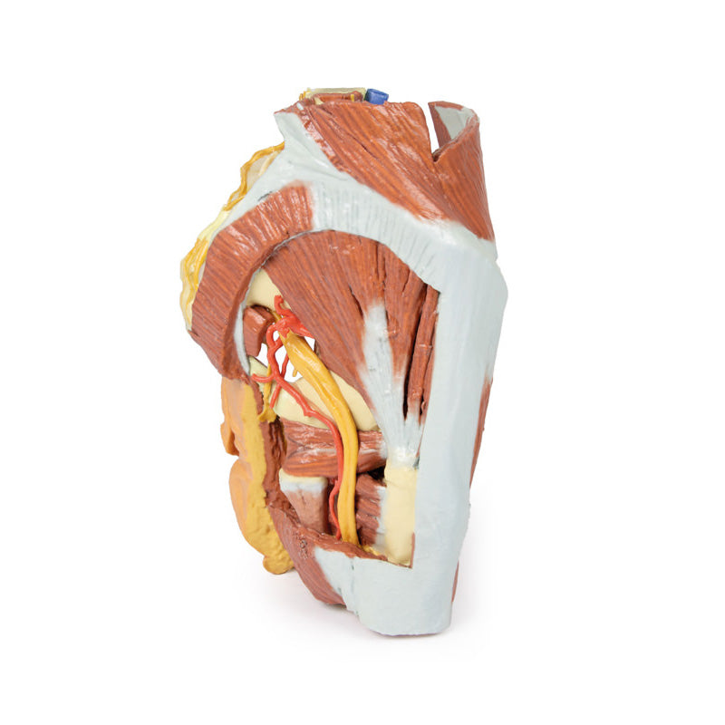
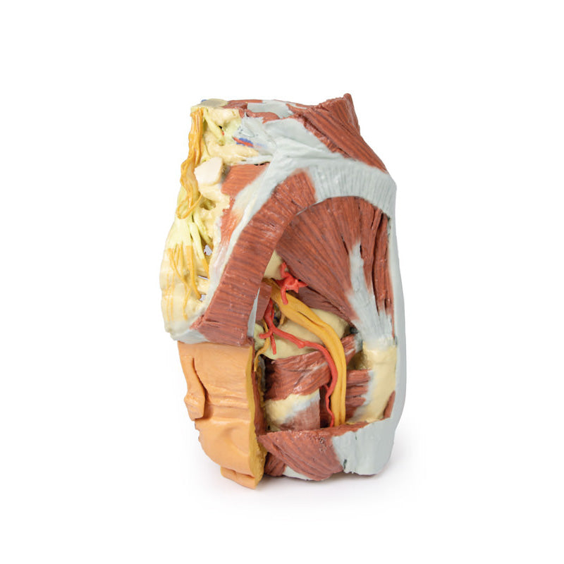
In the posterior view, most of the multifidus and origin of the gluteus maximus have beenremoved over the lumbr and sacral region, and the laminae of L4 and L5 and the dorsalsacrum have been sectioned to reveal the cauda equina in the vertebral and sacral canal.The dura mater has been partially sectioned to expose the roots traversing the region,including the passage of the sacral ventral rami through the ventral foramina. Laterally, alarge window into the gluteal maximus has been opened to expose the deeper structures ofthe gluteal region. Part of the sectioned piriformis is visible in the greater sciatic foramen,with the sciatic nerve (preserving an early division of the common peroneal and tibial nerveswithin the gluteal region) surrounded by the superior and inferior gluteal arteries. Thesectioned internal pudendal artery and pudendal nerve rest on the sacrotuberous ligamentas they descend towards the lesser sciatic foramen. Inferior to the sacrotuberous ligamentthe obturator internus muscle (along with the superior and inferior gemelli muscles) passeslaterally deep to the common peroneal and tibial nerves. Inferior to these lateral rotators,the quadratus femoris and common origin of the hamstring group are visible just proximalto the remaining portion of the gluteus maximus.
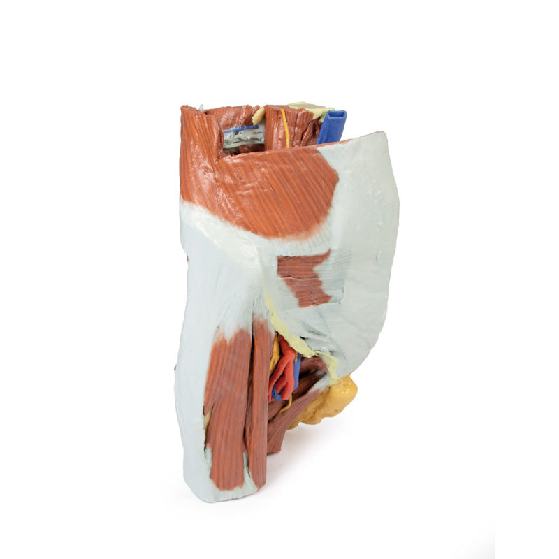
In the anterior view, a window has been cut into the aponeurosis of the external obliqueaponeurosis to reveal part of the transversus abdominis muscle. The inguinal ligament canbe seen arising from the anterior superior iliac spine and extending towards the pubictubercle. Inferior to the inguinal ligament, the fascia lata has been removed over theanterior thigh. The visible thigh muscles (from lateral to medial) on this specimen includethe tensor fasciae latae, and those of the anterior (sartorius, rectus femoris and theiliopsoas) and medial (gracilis, pectineus [sectioned], obturator externus, adductor brevis,adductor longus and adductor magnus).
Between these muscle groups the femoral sheath has been removed to expose the contentsof the femoral triangle (femoral artery and vein; sectioned to display the deeper adductormuscle fibers) and the femoral nerve that have entered this region deep to the inguinalligament. In this individual the lateral circumflex femoral artery arises directly from thefemoral artery. Inferior to this, the deep femoral artery (profunda femoris) branches off.Several anastomosing veins draining into the femoral vein surround the deep femoralartery. The femoral vein preserves an opening on the medial side corresponding to thedrainage point of the great saphenous vein. The medial circumflex artery, posterior branchof the obturator nerve and a muscular artery can be seen passing just superficial toobturator externus. The anterior branch of the obturator nerve can be seen more anteriorly,passing superficial to adductor magnus and deep to adductor longus.
The sectioned thigh of the specimen allows for orientation of muscles and neurovascularstructures in the proximal thigh. Other than the relations of anterior, medial and posteriorthigh muscles, perforating arteries and veins are visible near the adductor magnus; and thecommon peroneal and tibial nerves are visible in the posterior compartment.
Download Handling Guidelines for 3D Printed Models
GTSimulators by Global Technologies
Erler Zimmer Authorized Dealer

10.0 lb
🎄 HOLIDAY SAVINGS - Ends Dec 31 🎄
Discount has been automatically applied for this item.
3D Printed Female right pelvis
Item # MP1785
$3,048.00
$3,387.00
You save $339.00
Need an estimate?
Click Add To Quote

Features & Specifications
-
by
A trusted GT partner -
FREE Shipping
U.S. Contiguous States Only -
3D Printed Model
from a real specimen -
Gov't pricing
Available upon request
Frequently Bought Together
3D Printed Female right pelvis
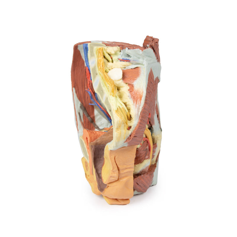
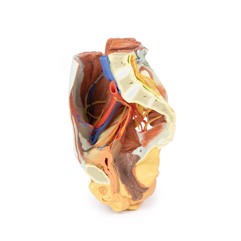
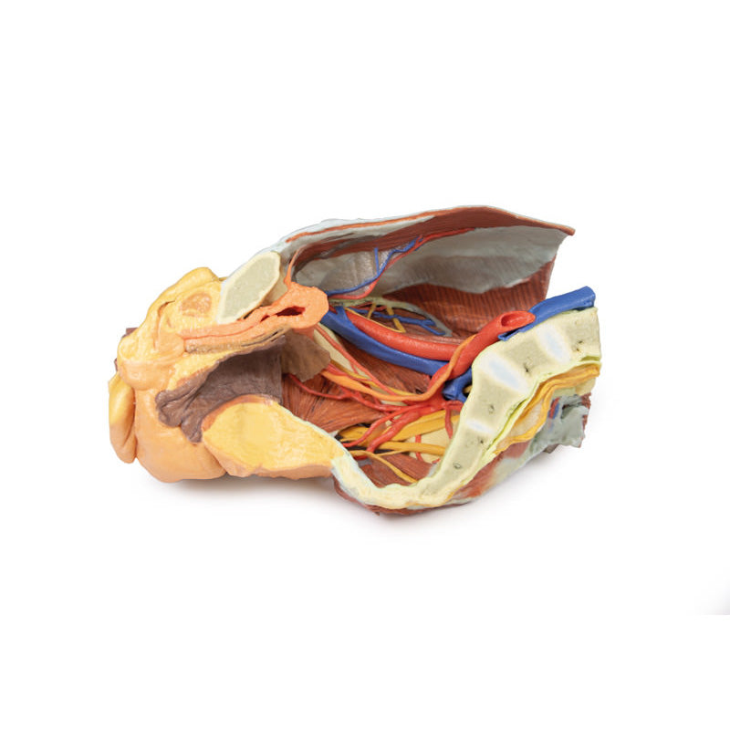
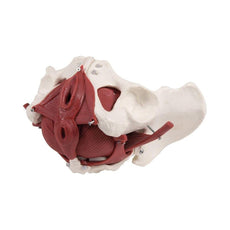
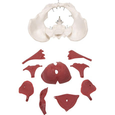
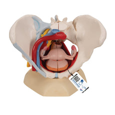
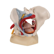
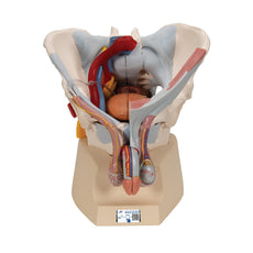
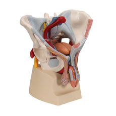
This 3D printed specimen represents a female right pelvis, sectioned along the midsagittalplane and transversely across the level of the L4 vertebrae and the proximal thigh. Thespecimen has been dissected to demonstrate the deep structures of the true and falsepelves, the inferior anterior abdominal wall and inguinal region, femoral triangle and glutealregion.Internal anatomy:
The muscular boundaries of the inferior abdominal cavity are defined posterolaterally bythe quadratus lumborum, iliacus and psoas muscles; anteriorly by the (varyinglyexposed) external and internal abdominal oblique muscles, the transversus abdominis andrectus abdominis. Inferiorly in the pelvic cavity, the obturator internus is visible traversingthrough the lesser sciatic foramen inferior to the sacrospinous ligament. Fibres of coccygeusmerge with those of the sacrospinous ligament. Piriformis has been sectioned, with bothorigin visible within the cavity (and part visible in the gluteal region). The common iliac artery arises from its cut edge at the level of L5, bifurcating at the level ofthe sacral promontory into the external and internal iliac arteries.
The external iliac arterycrosses the pelvic brim to give off the deep circumflex iliac artery and inferior epigastricartery before exiting the pelvis deep to the inguinal ligament. The internal iliac artery runsposterolaterally, giving the iliolumbar artery posteriorly and lateral sacral arteries whichenter the anterior sacral foramina.
A radicular artery entering the anterior foramina of thecoccyx can also be seen. Inferiorly, the superior gluteal artery, inferior gluteal artery andinternal pudendal artery exit the pelvic cavity through the greater sciatic foramen. A branchfrom the inferior gluteal artery, supplying psoas, travels anteriorly along the pectineal line.Anteriorly, the umbilical artery gives off the superior vesical artery (supplying the bladder)before terminating against the anterior abdominal wall as the medial umbilical ligament. Posteriorly, the inferior vesical artery arises from the obturator artery beforeexiting the pelvis through the obturator canal. The uterine artery crosses over the ureter toenter the remnants of the broad ligament.
The major veins preserved are the inferior epigastric vein and deep circumflex iliac veindraining into the external iliac vein, and the iliolumbar vein and lateral sacral vein draininginto the internal iliac vein. The external iliac vein and internal iliac vein unite to form theright common iliac vein which, at the level of L5, joins the (cut edge) of the left common iliacvein to become the inferior vena cava. Two veins pass horizontally across iliacus andquadratus lumborum.
Of the peripheral nerve structures preserved in this specimen, the lateral femoral cutaneousnerve passes laterally across the superficial aspect of the iliacus muscle and the femoralnerve is visible deep to the psoas major muscle. The genitofemoral nerve lies on thesuperficial surface of the psoas major, and the course of the genital branch entering thedeep inguinal ring and the femoral branch passing deep to the inguinal ligament can befollowed. The obturator nerve is also seen passing from deep to the psoas muscle anteriorlyto the obturator foramen. In the true pelvis the lumbosacral trunk crosses the pelvic brimand joints the anterior ramus of S1.
The S1-S3 anterior rami are visible and can be followedas they pass through the greater sciatic foramen and enter the gluteal region. In addition to the muscular and neurovascular structures, parts of pelvic viscera have beenpreserved. Posterior to the pubic symphysis the median umbilical ligament extendssuperiorly from the bladder to the anterior abdominal wall. The ureter descends anteriorlyto the psoas major muscle, across the iliac vessels, and beneath the uterine artery to enterthe posterior bladder wall. The urethra is seen passing downwards to its opening at theurinary meatus, just posterior to the clitoris. Posterior to the bladder are the posthysterectomyremnants of the uterus at the apex of the closed, superior end of the vagina.Posterior to this part of the cut rectum, anal canal and anus are present. Some muscularfibres of levator ani and the external anal sphincter can be seen in the ischioanal fossa justposteriorly to the anal canal.
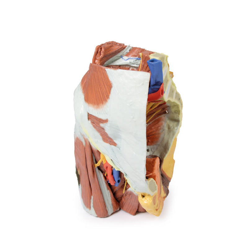
External anatomy:
In the posterior view, most of the multifidus and origin of the gluteus maximus have beenremoved over the lumbr and sacral region, and the laminae of L4 and L5 and the dorsalsacrum have been sectioned to reveal the cauda equina in the vertebral and sacral canal.The dura mater has been partially sectioned to expose the roots traversing the region,including the passage of the sacral ventral rami through the ventral foramina. Laterally, alarge window into the gluteal maximus has been opened to expose the deeper structures ofthe gluteal region. Part of the sectioned piriformis is visible in the greater sciatic foramen,with the sciatic nerve (preserving an early division of the common peroneal and tibial nerveswithin the gluteal region) surrounded by the superior and inferior gluteal arteries. Thesectioned internal pudendal artery and pudendal nerve rest on the sacrotuberous ligamentas they descend towards the lesser sciatic foramen. Inferior to the sacrotuberous ligamentthe obturator internus muscle (along with the superior and inferior gemelli muscles) passeslaterally deep to the common peroneal and tibial nerves. Inferior to these lateral rotators,the quadratus femoris and common origin of the hamstring group are visible just proximalto the remaining portion of the gluteus maximus.
In the anterior view, a window has been cut into the aponeurosis of the external obliqueaponeurosis to reveal part of the transversus abdominis muscle. The inguinal ligament canbe seen arising from the anterior superior iliac spine and extending towards the pubictubercle. Inferior to the inguinal ligament, the fascia lata has been removed over theanterior thigh. The visible thigh muscles (from lateral to medial) on this specimen includethe tensor fasciae latae, and those of the anterior (sartorius, rectus femoris and theiliopsoas) and medial (gracilis, pectineus [sectioned], obturator externus, adductor brevis,adductor longus and adductor magnus).
Between these muscle groups the femoral sheath has been removed to expose the contentsof the femoral triangle (femoral artery and vein; sectioned to display the deeper adductormuscle fibers) and the femoral nerve that have entered this region deep to the inguinalligament. In this individual the lateral circumflex femoral artery arises directly from thefemoral artery. Inferior to this, the deep femoral artery (profunda femoris) branches off.Several anastomosing veins draining into the femoral vein surround the deep femoralartery. The femoral vein preserves an opening on the medial side corresponding to thedrainage point of the great saphenous vein. The medial circumflex artery, posterior branchof the obturator nerve and a muscular artery can be seen passing just superficial toobturator externus. The anterior branch of the obturator nerve can be seen more anteriorly,passing superficial to adductor magnus and deep to adductor longus.
The sectioned thigh of the specimen allows for orientation of muscles and neurovascularstructures in the proximal thigh. Other than the relations of anterior, medial and posteriorthigh muscles, perforating arteries and veins are visible near the adductor magnus; and thecommon peroneal and tibial nerves are visible in the posterior compartment.
Download Handling Guidelines for 3D Printed Models
GTSimulators by Global Technologies
Erler Zimmer Authorized Dealer

by — Item # MP1785
3D Printed Female right pelvis
$3,048.00
$3,387.00
Add to Cart
Add to Quote


