Your shopping cart is empty.
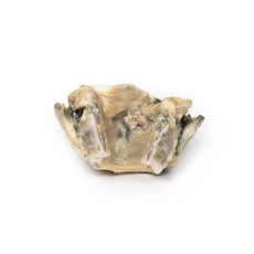
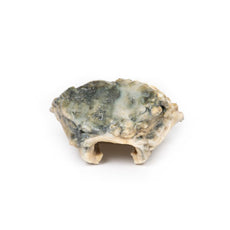
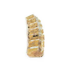
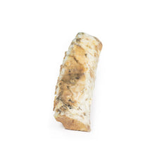
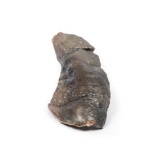
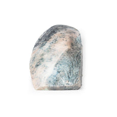
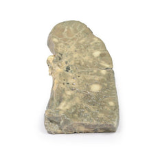
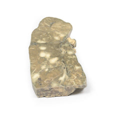
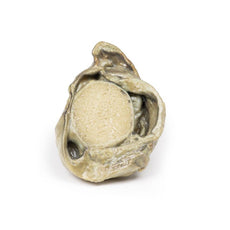
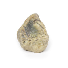
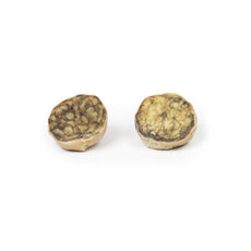
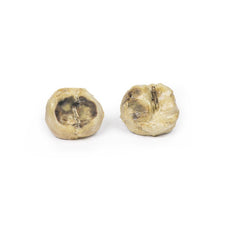
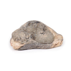
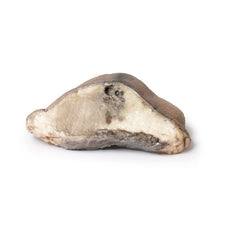
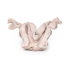
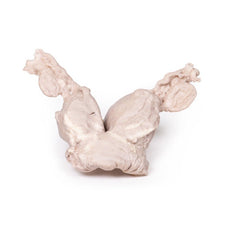
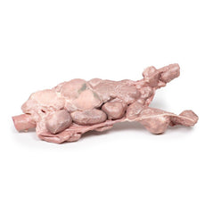
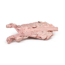
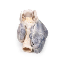
3D Printed Ventriculitis, Secondary to Septicaemia
Item # MP2008Need an estimate?
Click Add To Quote

-
by
A trusted GT partner -
3D Printed Model
from a real specimen -
Gov't pricing
Available upon request
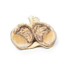
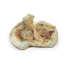
3D Printed Ventriculitis, Secondary to Septicaemia
Clinical History
A 50-year-old alcoholic was admitted with a 2-week history of weakness and shortness of
breath. At the onset of the illness he reported a productive cough, chest pain and blood-stained sputum.
Examination revealed a febrile, cyanosed, drowsy man with grunting respiration. A friction rub was present over
the right lower lobe. The remainder of the examination was unremarkable. The patient steadily deteriorated and
on the morning of his death, a lumbar puncture was performed. Green opalescent fluid was obtained. A blood
culture grew Streptococcus pneumoniae.
Pathology
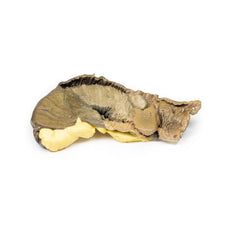
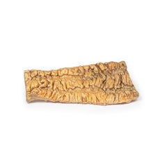
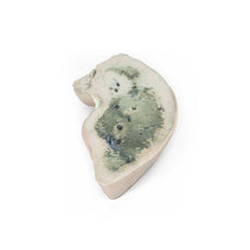
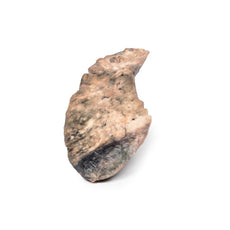
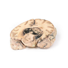
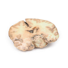
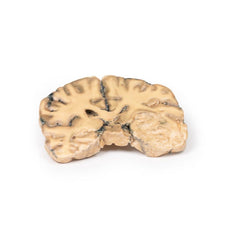
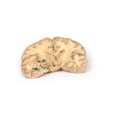
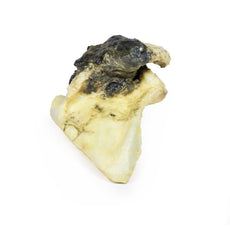
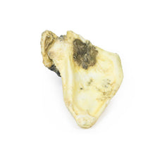
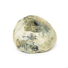
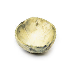
This specimen is an example of ventriculitis, with pneumococcal meningitis and right basal
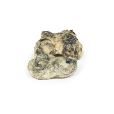
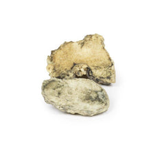
pneumonia also being found at autopsy. The horizontal slice through both cerebral hemispheres displays both of
the lateral ventricles. The ventricles show a thickened, rough ependymal lining with cellular debris
accumulation around the choroid plexus and also in the anterior horn. The lower surface shows similar changes
and also displays the normal arrangement of the caudate nucleus, lentiform nucleus and internal
capsule.
Histology demonstrated extensive infiltration of neutrophils in the sub-arachnoid space as well as
multifocal severe (sub)endothelial infiltration with obstruction of vascular lumen and involvement of the blood
vessel walls. The inflammation extended into the cerebral parenchyma causing haemorrhage and necrosis.
Further information
Ventriculitis is an uncommon complication of intracranial infection. In adults, it more
commonly occurs as a secondary complication of surgical intervention / instrumentation or trauma, rather than
from primary community-acquired meningitis. In these cases, causative organisms are similar to other nosocomial
(hospital-acquired) infections, in particular staphylococci or resistant Gram-negative bacilli. Neonates aged
less than 6 months have a higher incidence of ventricular infection. Presentation may be more subtle than in
bacterial meningitis or may be as obstructive hydrocephalus, secondary to resulting aqueductal obstruction.
Diagnosis is dependent on laboratory CSF testing and imaging, in particular using CT scans and MRI. Prolonged
intravenous antibiotic therapy is a mainstay of treatment with consideration to achieving effective
concentrations in CSF and brain tissue.
 Handling Guidelines for 3D Printed Models
GTSimulators by Global Technologies
Erler Zimmer Authorized Dealer
The models are very detailed and delicate. With normal production machines you cannot realize such details like shown in these models.
The printer used is a color-plastic printer. This is the most suitable printer for these models.
The plastic material is already the best and most suitable material for these prints. (The other option would be a kind of gypsum, but this is way more fragile. You even cannot get them out of the printer without breaking them).The huge advantage of the prints is that they are very realistic as the data is coming from real human specimen. Nothing is shaped or stylized.
The users have to handle these prints with utmost care. They are not made for touching or bending any thin nerves, arteries, vessels etc. The 3D printed models should sit on a table and just rotated at the table.


