Your shopping cart is empty.
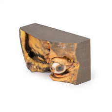
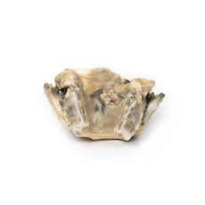
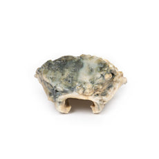
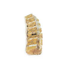
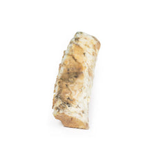
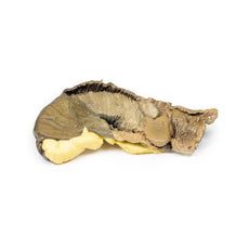
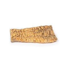
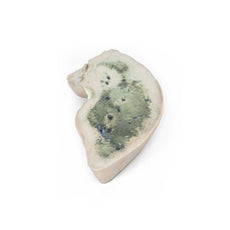
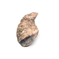
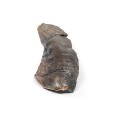
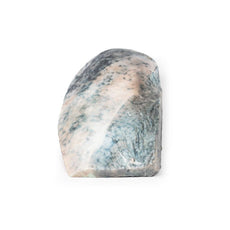
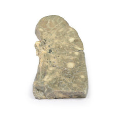
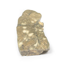
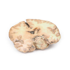
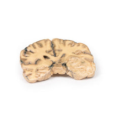
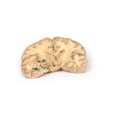
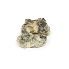
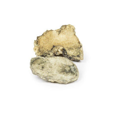
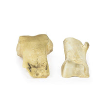
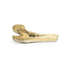
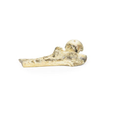
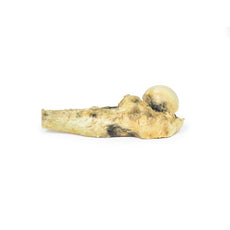
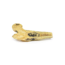
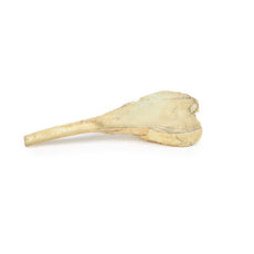
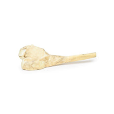
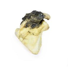
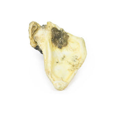
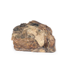
3D Printed Hilum of the Right Lung
Item # MP1127Need an estimate?
Click Add To Quote

-
by
A trusted GT partner -
FREE Shipping
U.S. Contiguous States Only -
3D Printed Model
from a real specimen -
Gov't pricing
Available upon request
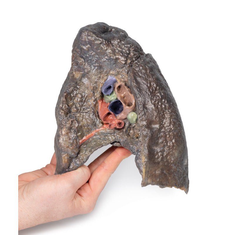
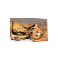
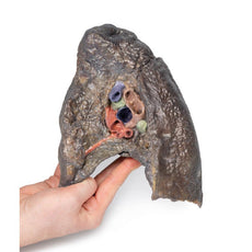
3D Printed Hilum of the Right Lung
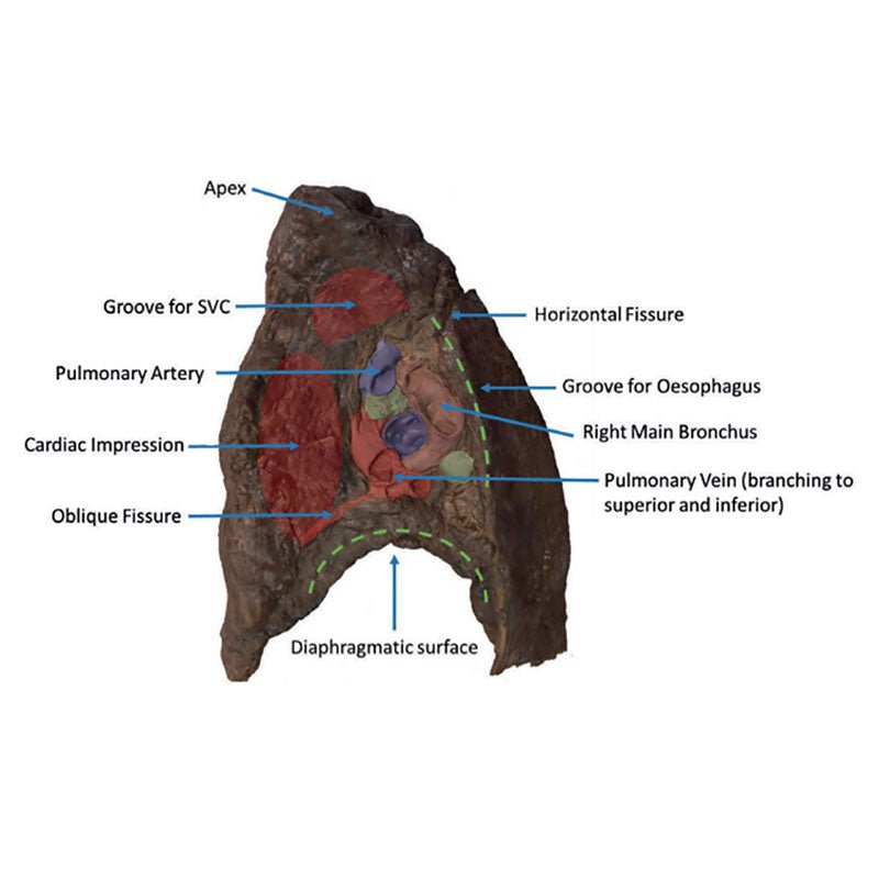
The hilum of a lung is the point at which visceral and parietal pleura meet and functions with the pulmonary
ligament as the lungs only connection with the rest of the body. This connection includes the Pulmonary Artery,
Superior and Inferior Pulmonary Veins, Main Bronchi, Nerves and Lymphatics.
As the definition of an artery
involves carrying blood AWAY from the heart, this will be deoxygenated blood in the pulmonary system, in
contrast with the systemic circulation. Similarly, veins carry blood TOWARDS the heart, meaning it will be
oxygenated in the pulmonary system.
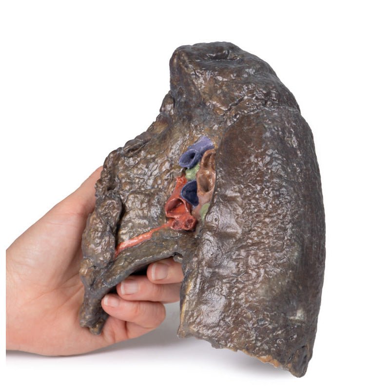
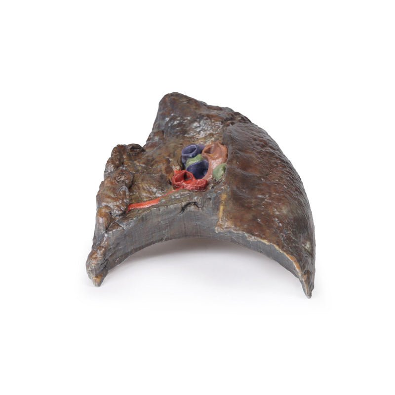
With the specimen cut in a sagittal plane in line with the cardiac impression, nerves and lymphatics are difficult to identify however the groove from the oesophagus as it descends posteriorly to pierce the diaphragm can be seen alongside the cardiac impression (of the right atrium) is notable anterior to the hilum of the right lung; the right main bronchi and its subsequent divisions into lobar bronchi, found in this specimen more posterior in the hilum; he pulmonary artery and its divisions, located most superior within the hilum; the superior and inferior pulmonary veins and their divisions which are most inferior and anterior in the specimen. the oblique and horizontal fissures along the lateral surface of the specimen and the Hilar lymph nodes around the hilum on the medial surface of the lung.
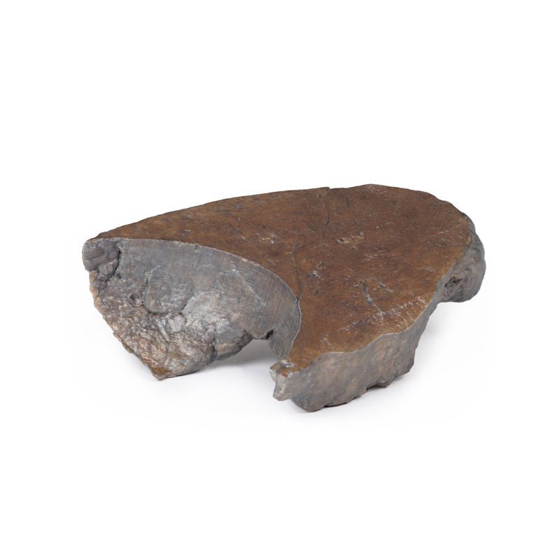
The diaphragmatic surface is found inferiorly and the costal visceral surface is on the posterior of the specimen.
Download: Handling Guidelines for 3D Printed Models
GTSimulators by Global Technologies
Erler Zimmer Authorized Dealer
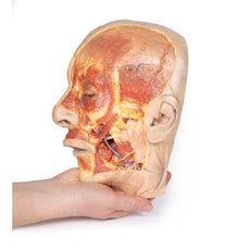
The models are very detailed and delicate. With normal production machines you cannot realize such details like shown in these models.
The printer used is a color-plastic printer. This is the most suitable printer for these models.
The plastic material is already the best and most suitable material for these prints. (The other option would be a kind of gypsum, but this is way more fragile. You even cannot get them out of the printer without breaking them).The huge advantage of the prints is that they are very realistic as the data is coming from real human specimen. Nothing is shaped or stylized.
The users have to handle these prints with utmost care. They are not made for touching or bending any thin nerves, arteries, vessels etc. The 3D printed models should sit on a table and just rotated at the table.


