Your shopping cart is empty.
3D Printed Cerebral Haemorrhage
Item # MP2002Need an estimate?
Click Add To Quote

-
by
A trusted GT partner -
3D Printed Model
from a real specimen -
Gov't pricing
Available upon request
3D Printed Cerebral Haemorrhage
Clinical History
A woman of 56-years was admitted following 2 episodes of severe headache with loss of
consciousness. Clinical examination revealed systemic hypertension with cardiac enlargement, and a right
hemiparesis. Angiography showed bilateral middle cerebral aneurysms. The patient‘s condition deteriorated, and she
died soon after admission.
Pathology
The left hemispher
3D Printed Cerebral Haemorrhage
Clinical History
A woman of 56-years was admitted following 2 episodes of severe headache with loss of
consciousness. Clinical examination revealed systemic hypertension with cardiac enlargement, and a right
hemiparesis. Angiography showed bilateral middle cerebral aneurysms. The patient‘s condition deteriorated, and she
died soon after admission.
Pathology
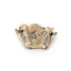
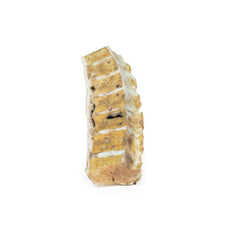
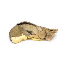
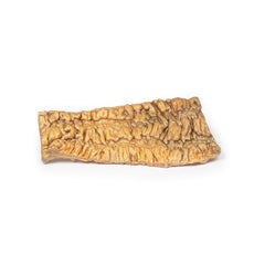
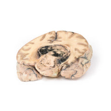
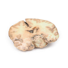
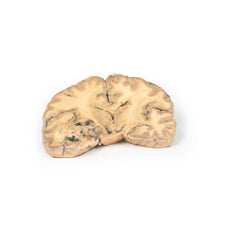
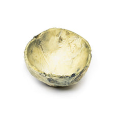
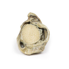
The left hemisphere of the brain has been sliced in the parasagittal plane, and the cut surface displays
a large cerebral haemorrhage in the parietal and frontal lobe. The haemorrhage and associated clot are causing
extensive distortion of the left external capsule and lateral ventricle. The source of the bleeding was a ruptured
aneurysm of the left middle cerebral artery.
 Handling Guidelines for 3D Printed Models
GTSimulators by Global Technologies
Erler Zimmer Authorized Dealer
The models are very detailed and delicate. With normal production machines you cannot realize such details like shown in these models.
The printer used is a color-plastic printer. This is the most suitable printer for these models.
The plastic material is already the best and most suitable material for these prints. (The other option would be a kind of gypsum, but this is way more fragile. You even cannot get them out of the printer without breaking them).The huge advantage of the prints is that they are very realistic as the data is coming from real human specimen. Nothing is shaped or stylized.
The users have to handle these prints with utmost care. They are not made for touching or bending any thin nerves, arteries, vessels etc. The 3D printed models should sit on a table and just rotated at the table.


