Your shopping cart is empty.
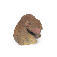
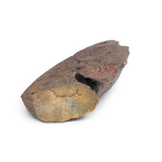
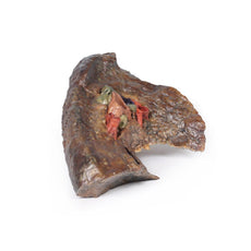
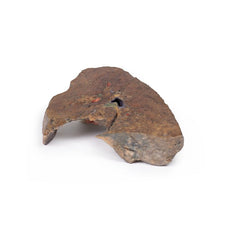
3D Printed Heart
Item # MP1123Need an estimate?
Click Add To Quote

-
by
A trusted GT partner -
FREE Shipping
U.S. Contiguous States Only -
3D Printed Model
from a real specimen -
Gov't pricing
Available upon request
3D Printed Heart
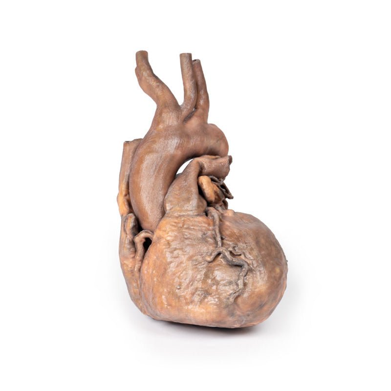
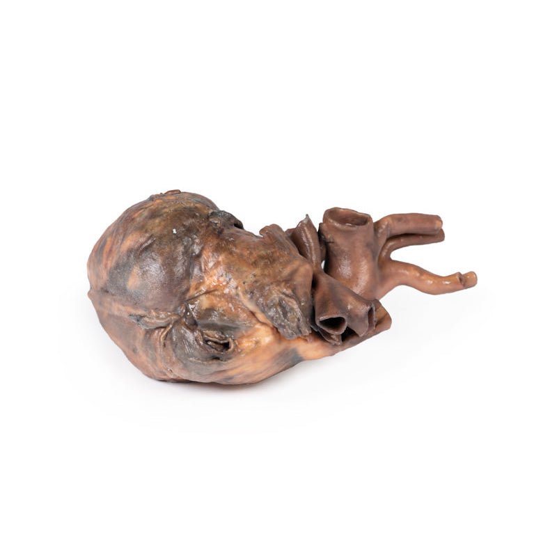
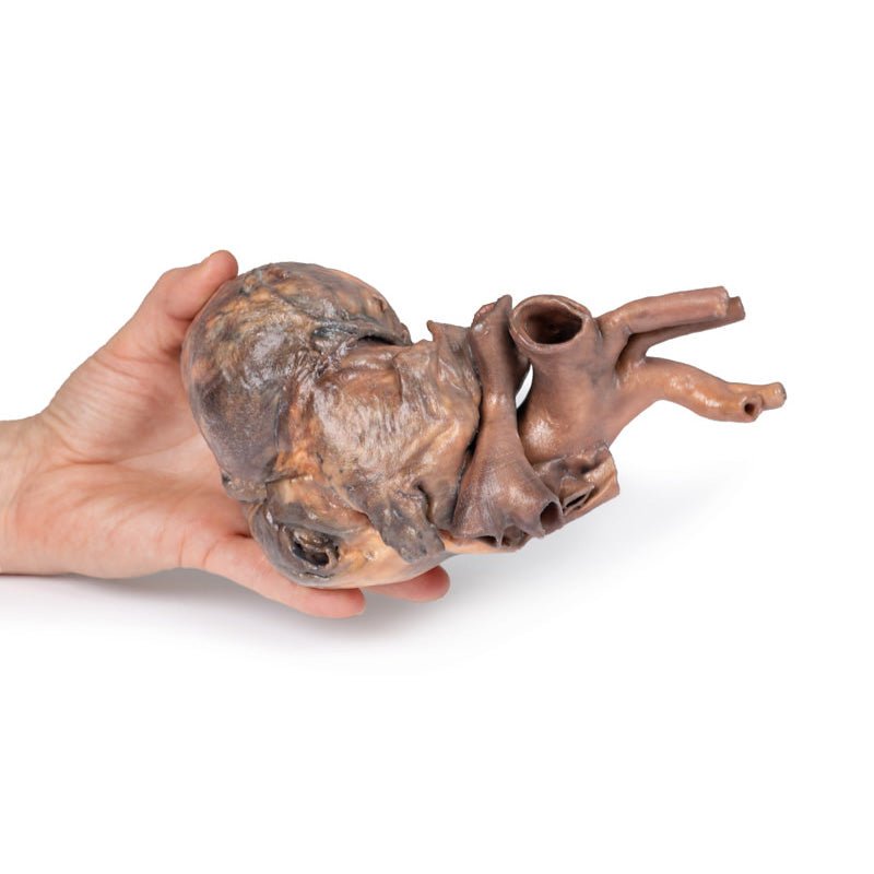
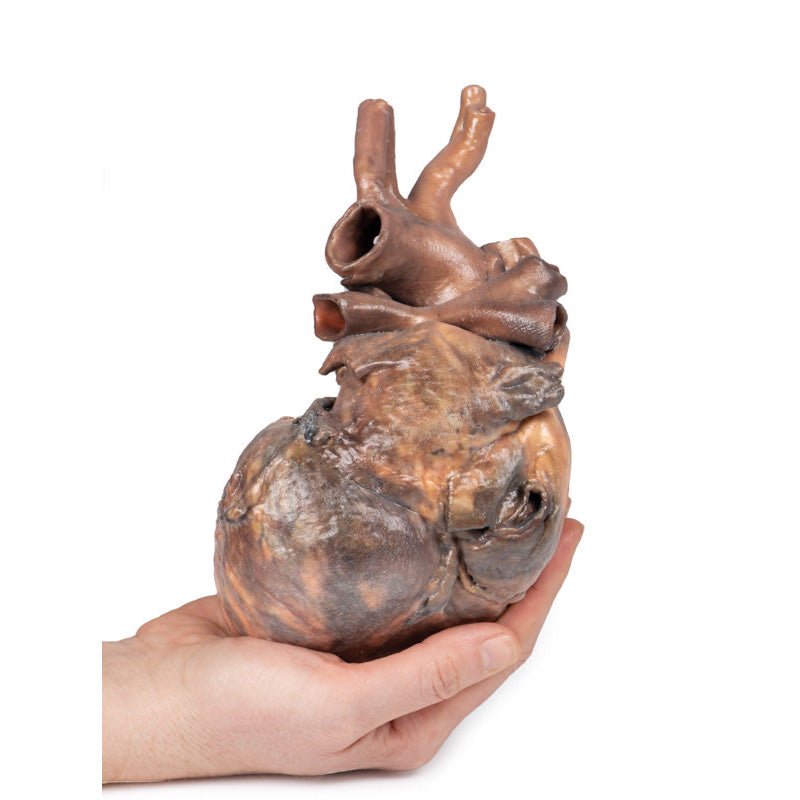
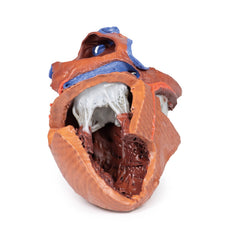
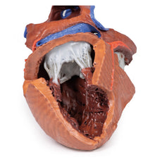
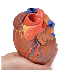
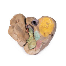
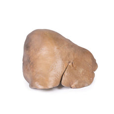
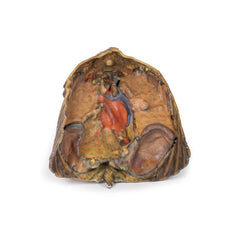
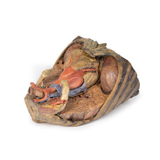
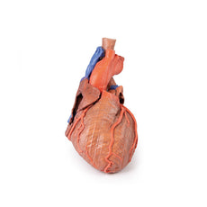
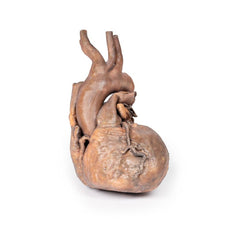
This 3D model represents a ‘normal’ sized adult heart with light dissection
to the epicardium to expose the coronary arteries and cardiac veins.
At the base of the heart, the terminal part of the superior vena cava and
azygous vein can be observed just prior to draining into the right atrium.
Immediately adjacent to the superior vena cava, the arch of the aorta has
been preserved with the origins of the ao
3D Printed Heart
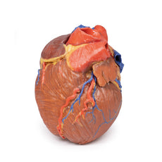
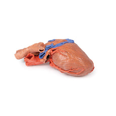
This 3D model represents a ‘normal’ sized adult heart with light dissection
to the epicardium to expose the coronary arteries and cardiac veins.
At the base of the heart, the terminal part of the superior vena cava and
azygous vein can be observed just prior to draining into the right atrium.
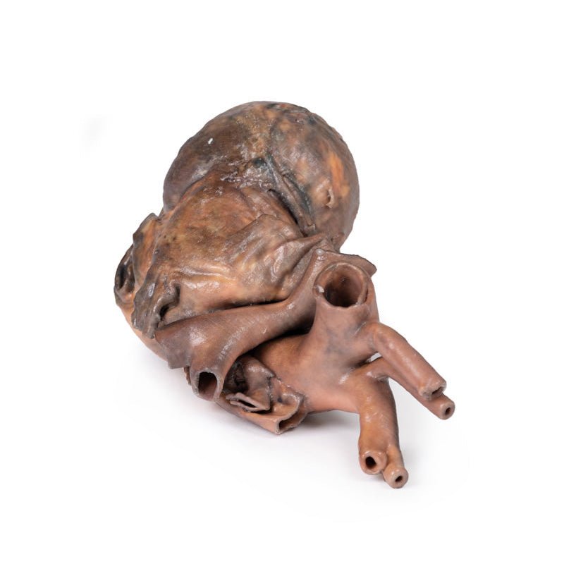
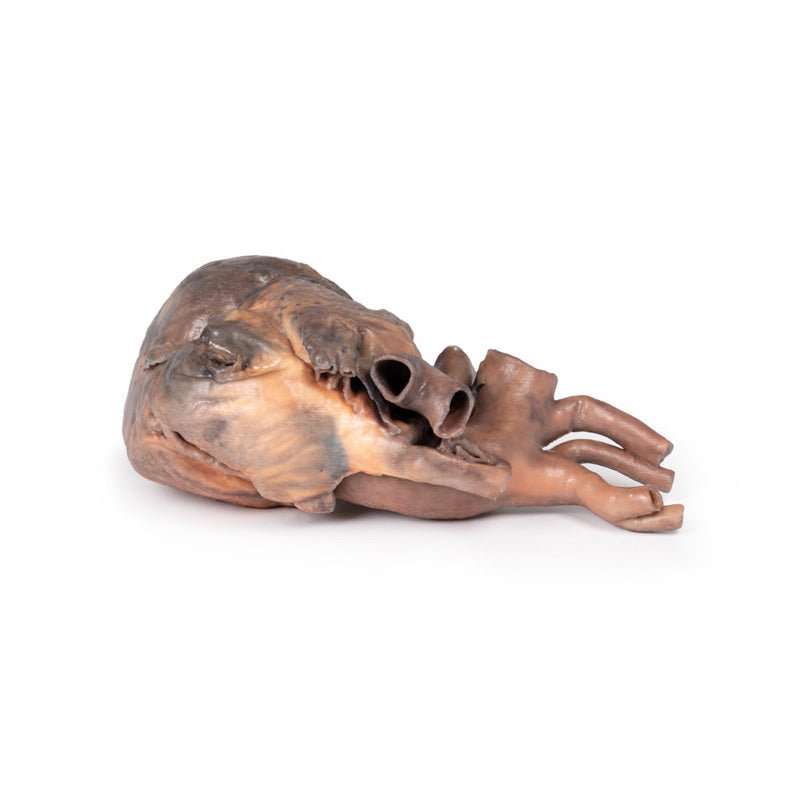
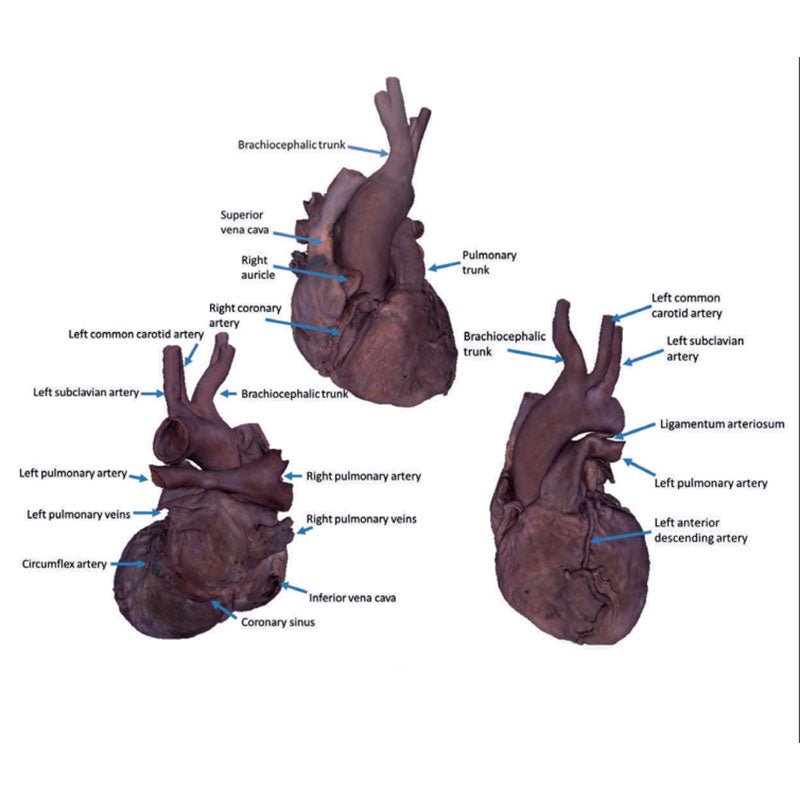
Immediately adjacent to the superior vena cava, the arch of the aorta has
been preserved with the origins of the aortic arch derivative arteries. In
slight contrast to the typical branching pattern, the brachiocephalic trunk
includes both the right subclavian and right common carotids as well as
the left common carotid. As a result, only two direct arterial branches
can be observed – with the unusually joined brachiocephaic trunk and
left subclavian arising before the descent of the thoracic aorta posterior
to the pulmonary trunk. The pulmonary trunk and pulmonary arteries are
preserved, including a robust ligamentum arteriosum connecting the left
pulmonary artery to the aortic arch.
The removal of the epicardium has exposed the coronary arteries and
branches across both the anterior and posterior aspects of the heart
chambers. The right coronary artery can be seen descending from its
origin at the ascending aorta, and wrapping posteriorly to approach the
posterior interventricular sulcus. The origin of the left coronary artery
is obscured by the auricle of the left atrium, but the branches from this
artery – the anterior interventricular (left anterior descending), diagonal
(passing deep into the myocardium) and the circumflex artery can
be observed at the superior margin of the left ventricle. The anterior
interventricular descends towards the apex with several branches passing
deep into the myocardium, while the circumflex passes posteriorly and
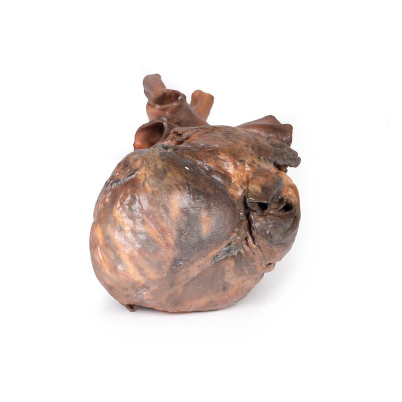
lies just superficial to a preserved portion of the great cardiac vein. On
the posterior aspect, a well-defined coronary sinus is preserved to its
termination at the right atrium near the inferior vena cava.
 Handling Guidelines for 3D Printed Models
GTSimulators by Global Technologies
Erler Zimmer Authorized Dealer
The models are very detailed and delicate. With normal production machines you cannot realize such details like shown in these models.
The printer used is a color-plastic printer. This is the most suitable printer for these models.
The plastic material is already the best and most suitable material for these prints. (The other option would be a kind of gypsum, but this is way more fragile. You even cannot get them out of the printer without breaking them).The huge advantage of the prints is that they are very realistic as the data is coming from real human specimen. Nothing is shaped or stylized.
The users have to handle these prints with utmost care. They are not made for touching or bending any thin nerves, arteries, vessels etc. The 3D printed models should sit on a table and just rotated at the table.


