Your shopping cart is empty.
3D Printed Horseshoe Kidney
Item # MP2092Need an estimate?
Click Add To Quote

-
by
A trusted GT partner -
FREE Shipping
U.S. Contiguous States Only -
3D Printed Model
from a real specimen -
Gov't pricing
Available upon request
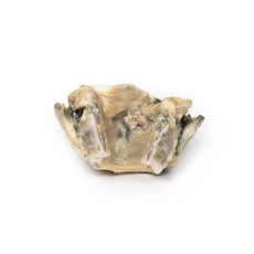
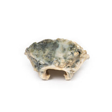
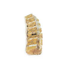
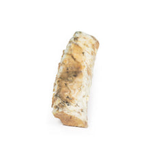
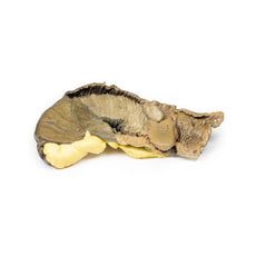
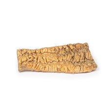
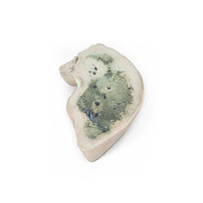
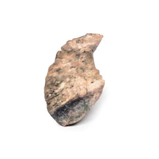
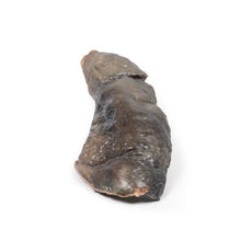
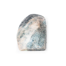
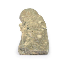
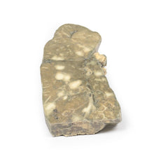
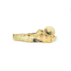
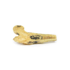
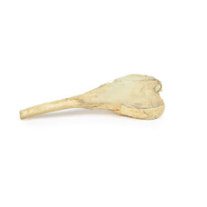
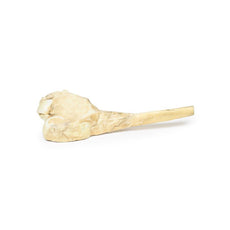
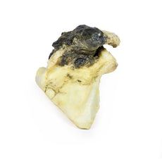
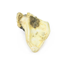
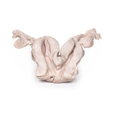
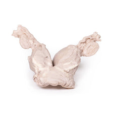
3D Printed Horseshoe Kidney
Clinical History
This specimen was found during a routine post-mortem of a 56-year-old male who
died of rheumatic heart disease.
Pathology
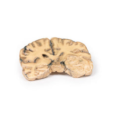
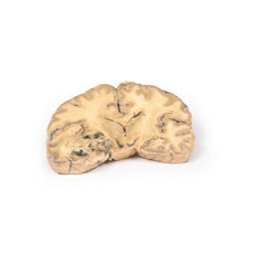
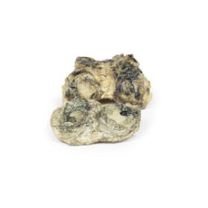
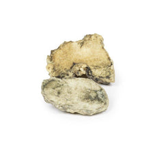
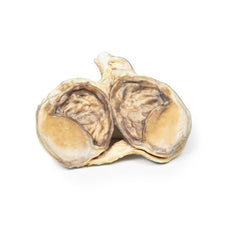
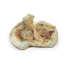
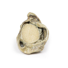
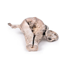
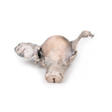
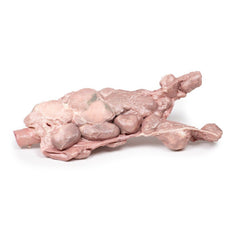
The kidney is 12 cm in length and the two parts are fused at the lower pole forming
this horseshoe-like shape. The ureters can be seen emerging from the hilum on the anterior aspect of the 3D print.
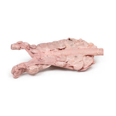
The kidney is bisected in the horizontal pl
3D Printed Horseshoe Kidney
Clinical History
This specimen was found during a routine post-mortem of a 56-year-old male who
died of rheumatic heart disease.
Pathology
The kidney is 12 cm in length and the two parts are fused at the lower pole forming
this horseshoe-like shape. The ureters can be seen emerging from the hilum on the anterior aspect of the 3D print.
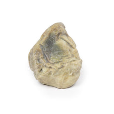
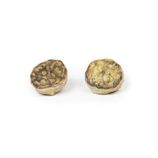
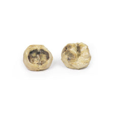
The kidney is bisected in the horizontal plane which is evident on the posterior aspect. There is persistent foetal
lobulation. The renal pelvis is characteristically antero-medial positioned with the ureters travelling anterior to
the fused lower poles or isthmus of the kidney.
Further Information
Horseshoe kidneys are the commonest renal developmental abnormality. This
anomaly is twice as common in males as in females. They are found in about 1 in 500 to 1000 post-mortem
examinations. Most cases are sporadic but may be associated with some chromosomal anomalies, such as those resulting
in Downs and Edwards Syndromes as well as non-aneuploidic anomalies, such as VACTERL* association. In 90% of cases
an isthmus of renal tissue connects the lower poles of the kidneys across the midline, forming a horseshoe shape.
Fusion of the upper lobes is rare. The renal pelves that drain the two halves of the horseshoe kidney are directed
more anteriorly than normal and there is some angulation of ureters as they cross the isthmus.
This malformation is usually asymptomatic and picked up incidentally on routine ultrasound or CT scans. These kidneys
usually function normally. There is an increased incidence of urinary calculi, possibly due to angulation of the
ureters and to the resulting stasis. There is an increased risk of hydronephrosis usually from pelvi-ureteric
junction obstruction. There is a higher incidence of urinary tract infections mainly due to vesico-ureteric reflux.
A higher incidence in some forms of renal malignancies (e.g. transitional cell carcinoma and Wilms tumours) has also
been described.
(*VACTERL = Vertebral defects, Anal atresia, Cardiac defects, Tracheo-Esophageal fistula, Renal
anomalies, and Limb abnormalities).
 Handling Guidelines for 3D Printed Models
GTSimulators by Global Technologies
Erler Zimmer Authorized Dealer
The models are very detailed and delicate. With normal production machines you cannot realize such details like shown in these models.
The printer used is a color-plastic printer. This is the most suitable printer for these models.
The plastic material is already the best and most suitable material for these prints. (The other option would be a kind of gypsum, but this is way more fragile. You even cannot get them out of the printer without breaking them).The huge advantage of the prints is that they are very realistic as the data is coming from real human specimen. Nothing is shaped or stylized.
The users have to handle these prints with utmost care. They are not made for touching or bending any thin nerves, arteries, vessels etc. The 3D printed models should sit on a table and just rotated at the table.


