Your shopping cart is empty.
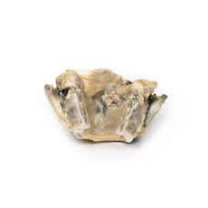
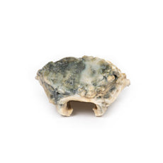
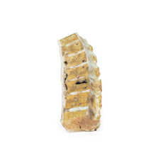
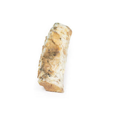
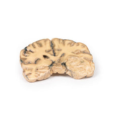
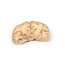
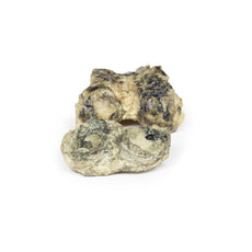
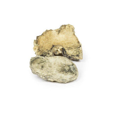
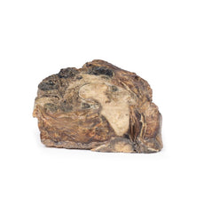
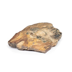
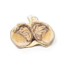
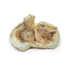
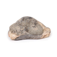
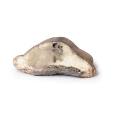
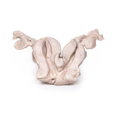
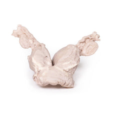
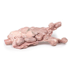
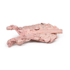
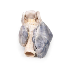
3D Printed Hypertrophic Subaortic Stenosis
Item # MP2036Need an estimate?
Click Add To Quote

-
by
A trusted GT partner -
FREE Shipping
U.S. Contiguous States Only -
3D Printed Model
from a real specimen -
Gov't pricing
Available upon request
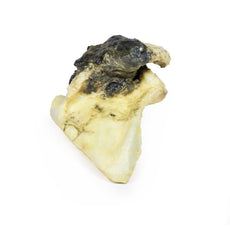
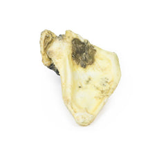
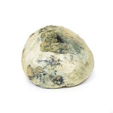
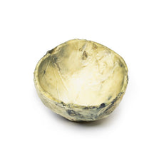
3D Printed Hypertrophic Subaortic Stenosis
Clinical History
A thin 42-year-old American tourist was found dead in his hotel bedroom. A coroner’s autopsy
was performed.
Pathology
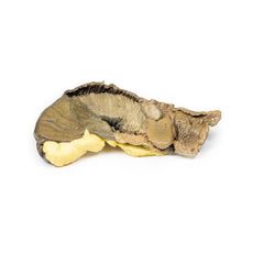
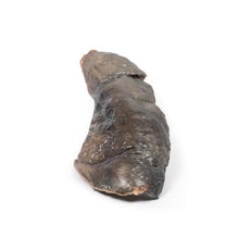
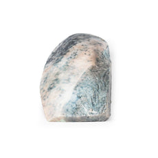
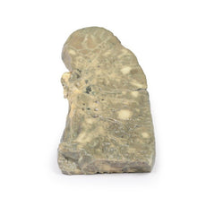
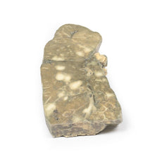
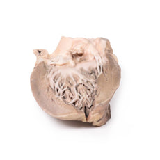
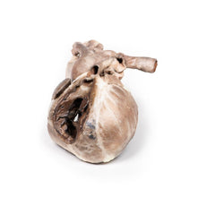
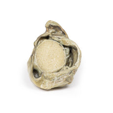
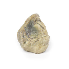
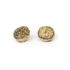
This is a longitudinal section through the heart displaying the left and right ventricles and
interventricular septum. The outstanding abnormality is a grossly thickened interventricular septum and left
ventricular hypertrophy. The aortic cusps that are visible appear unremarkable, as does the mitral valve. The
ventricular septum is so large that it encroaches on the lumen of the left ventricle.
Diagnosis
Idiopathic hypertrophic subaortic stenosis, also known as hypertrophic cardiomyopathy.
Further Information
Subaortic stenosis is considered to be acquired rather than congenital and is suggested to
result from an underlying defect in the architecture of the left ventricular outflow tract (LVOT). The defect
may be such that the resulting turbulent blood flow leads to progressive thickening and fibrosis of the LVOT and
the aortic valve. Progression of the disease will often lead to a hypertrophic cardiomyopathy secondary to the
increased aortic pressure needed to be overcome by the left ventricle. Mild or moderate stenosis is often
asymptomatic. As disease progresses and the stenosis becomes severe, symptoms such as exertional dyspnoea and
syncope may become apparent. Investigation and subsequent diagnosis are often prompted by the presence of an
ejection systolic murmur on physical examination. Echocardiography is used to confirm diagnosis.
These days,
definitive treatment of subaortic stenosis consists of surgical correction of the obstruction.
 Handling Guidelines for 3D Printed Models
GTSimulators by Global Technologies
Erler Zimmer Authorized Dealer
The models are very detailed and delicate. With normal production machines you cannot realize such details like shown in these models.
The printer used is a color-plastic printer. This is the most suitable printer for these models.
The plastic material is already the best and most suitable material for these prints. (The other option would be a kind of gypsum, but this is way more fragile. You even cannot get them out of the printer without breaking them).The huge advantage of the prints is that they are very realistic as the data is coming from real human specimen. Nothing is shaped or stylized.
The users have to handle these prints with utmost care. They are not made for touching or bending any thin nerves, arteries, vessels etc. The 3D printed models should sit on a table and just rotated at the table.


