Your shopping cart is empty.
3D Printed Metastatic Tumour in Lung from Primary Testicular Cancer
Item # MP2055Need an estimate?
Click Add To Quote

-
by
A trusted GT partner -
FREE Shipping
U.S. Contiguous States Only -
3D Printed Model
from a real specimen -
Gov't pricing
Available upon request
3D Printed Metastatic Tumour in Lung from Primary Testicular Cancer
Clinical History
A 37-year old male patient presents with a 1-month history of lethargy, cough
and weight loss. He had a history of an orchiectomy 18 months previous for a testicular tumour. Then 12 months
post-op he underwent neck radiotherapy to treat metastasis. On admission, he became acutely dyspnoeic and hypoxic
and died.
Pathology
T
3D Printed Metastatic Tumour in Lung from Primary Testicular Cancer
Clinical History
A 37-year old male patient presents with a 1-month history of lethargy, cough
and weight loss. He had a history of an orchiectomy 18 months previous for a testicular tumour. Then 12 months
post-op he underwent neck radiotherapy to treat metastasis. On admission, he became acutely dyspnoeic and hypoxic
and died.
Pathology
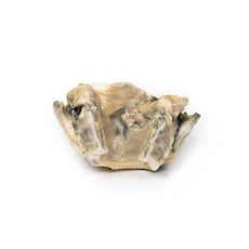
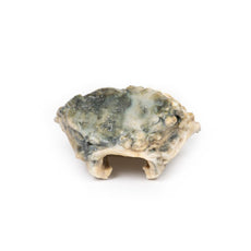
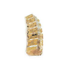
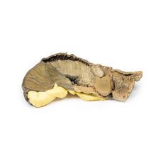
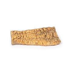
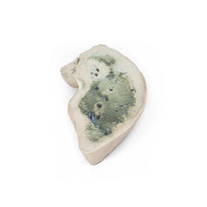
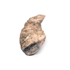
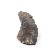
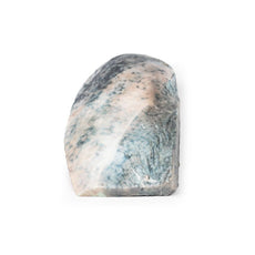
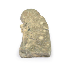
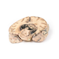
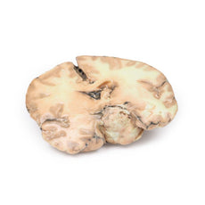
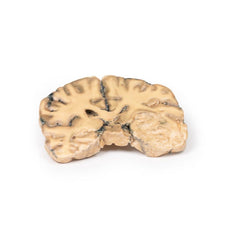
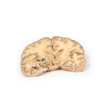
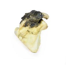
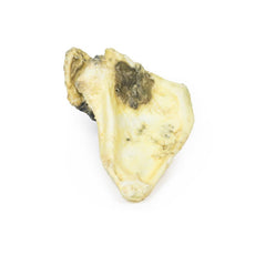
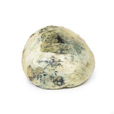
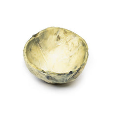
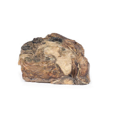
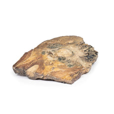
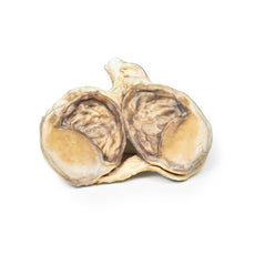
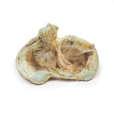
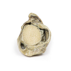
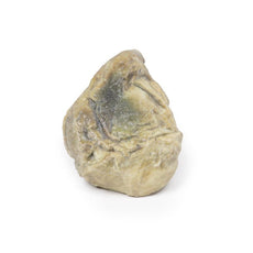
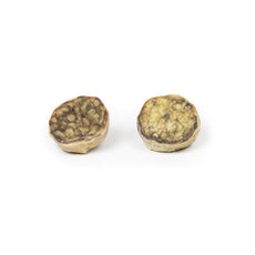
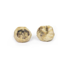
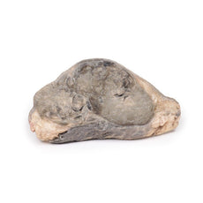
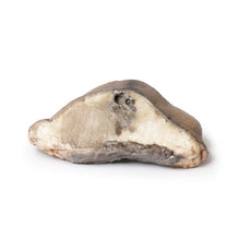
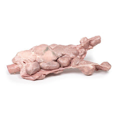
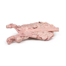
This right lung specimen (and portions of 4 ribs) has been sliced longitudinally.
There are numerous rounded tumour nodules evident in the lung parenchyma ranging from 5 to 30mm in diameter. The
tumours are variegated in appearance with pale yellow and dark brown cut surfaces. One tumour is extending along the
bronchus of the lower lobe forming a cast. Several nodules project from the pleural surface and some show central
umbilication from necrosis and haemorrhage. This is an example of pulmonary metastases from a mixed germ cell
testicular tumour, most likely choriocarcinoma arising in a malignant teratoma.
Further Information
Germ cell testicular tumours (GCT) are the most common tumours found in men.
Average age of diagnosis is 30 year of age and are rarely diagnosed pre-puberty. Risk factors for development
include cryptorchidism and a positive family history of GCT. Familial GCT increased risk can be linked to genes
encoding for kinases, e.g. KIT and BAK.
They can be divided into two groups: seminomatous (resemble primordial germ cells) and non-seminomatous (resemble embryonic stem cells). Over one third of GCT are mixed GCT, with two or more GCT types in one mass. Many possible combinations of seminoma, teratoma, embryonal carcinoma, yolk sac tumor, and choriocarcinoma can be seen. Teratomas components are found in one third of mixed GCT. Elevated serum Alpha Fetoprotein and beta-hCG are produced by choriocarcinoma. Lymphatic spread involves the retroperitoneal para aortic nodes initially. Mediastinal and supraclavicular nodes may later become involved. The lungs are the most common site for haematogenous spread but the liver, brain or bones may also be affected.
Symptoms may include a painless testicular mass and haematospermia. Later symptoms of distant metastases may occur.
Common symptoms of lung metastases include cough, dyspnoea, haemoptysis, recurrent infection
Treatment depends on
clinical stage but usually involves radical orchiectomy, chemotherapy and sometimes radiotherapy. More than 95% on
early stage GCT can be cured.
 Handling Guidelines for 3D Printed Models
GTSimulators by Global Technologies
Erler Zimmer Authorized Dealer
The models are very detailed and delicate. With normal production machines you cannot realize such details like shown in these models.
The printer used is a color-plastic printer. This is the most suitable printer for these models.
The plastic material is already the best and most suitable material for these prints. (The other option would be a kind of gypsum, but this is way more fragile. You even cannot get them out of the printer without breaking them).The huge advantage of the prints is that they are very realistic as the data is coming from real human specimen. Nothing is shaped or stylized.
The users have to handle these prints with utmost care. They are not made for touching or bending any thin nerves, arteries, vessels etc. The 3D printed models should sit on a table and just rotated at the table.


