Your shopping cart is empty.
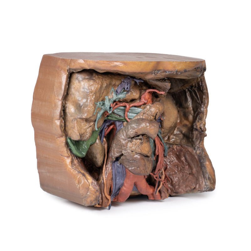
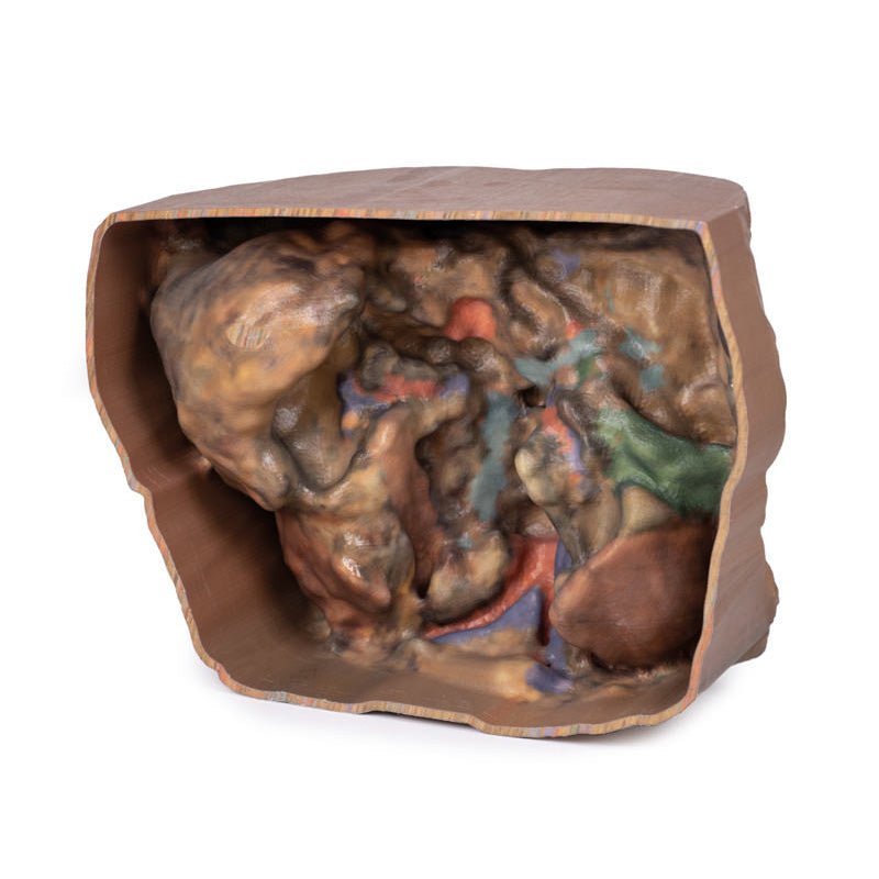
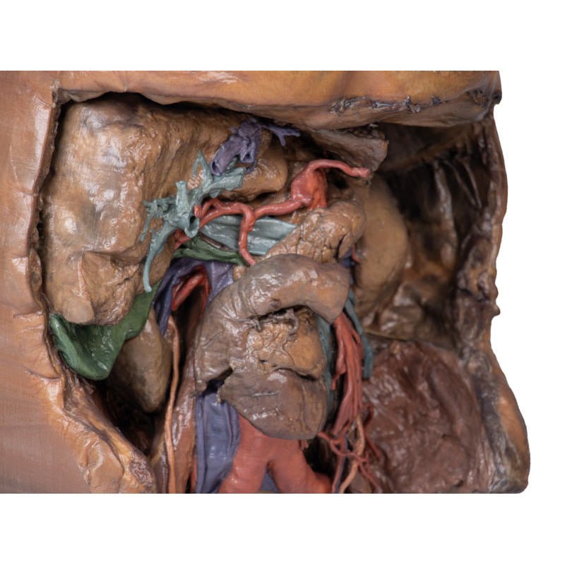
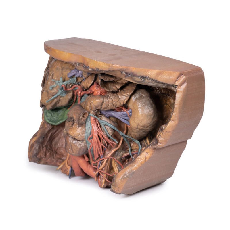
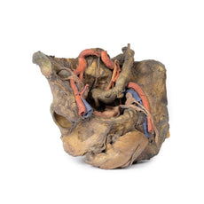
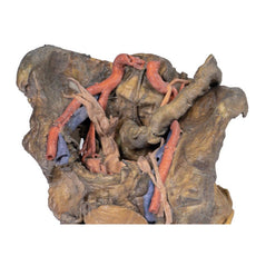
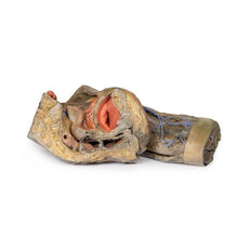
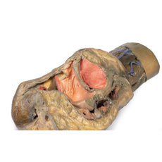
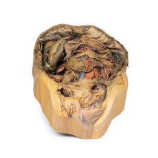
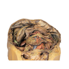
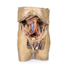
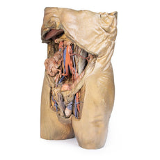
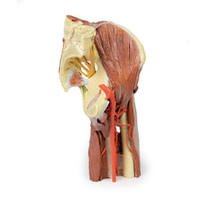
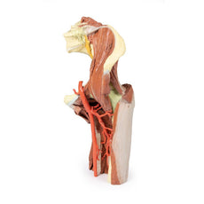
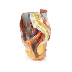
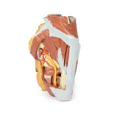
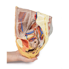
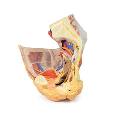
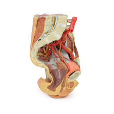
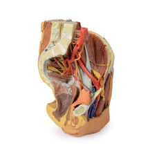
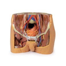
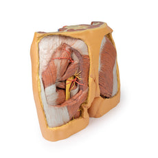
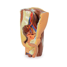
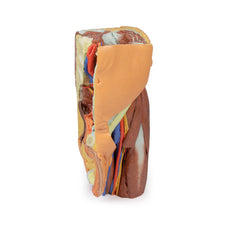
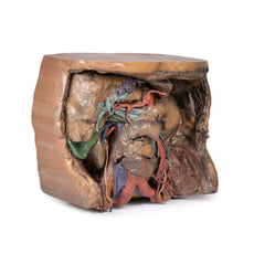
3D Printed Abdomen Vasculature
Item # MP1131Need an estimate?
Click Add To Quote

-
by
A trusted GT partner -
FREE Shipping
U.S. Contiguous States Only -
3D Printed Model
from a real specimen -
Gov't pricing
Available upon request
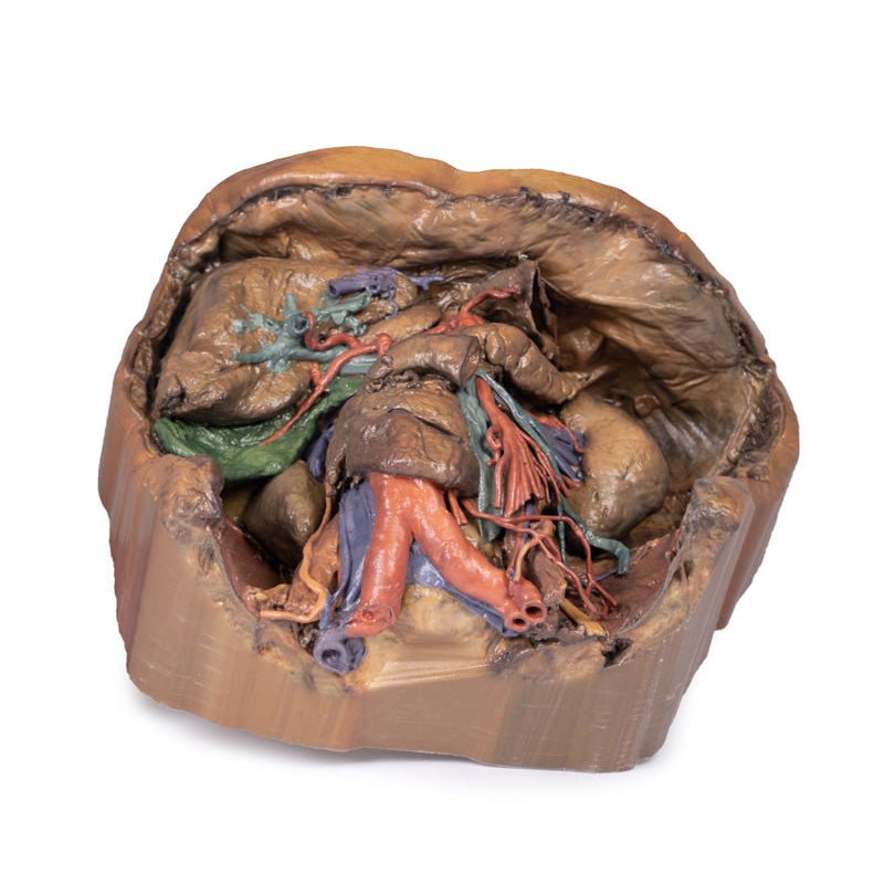
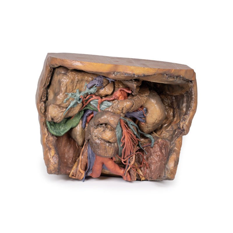
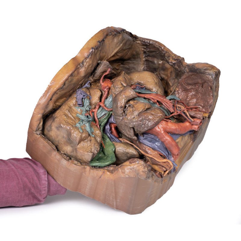
3D Printed Abdomen Vasculature
Coeliac Trunk
Supplying the embryological foregut, the celiac trunk arises from T12 spinal
level. Branches that can be observed in this specimen include the Left gastric artery arising from the left
portion of the celiac trunk; remains of the splenic artery arising from the celiac trunk and visible passing to
the left hypochondrium; the Common hepatic artery, located to the right of the celiac trunk and giving off key
branches; the Gastroduodenal artery, branching inferior to into the right gastric artery, and provide an
anastomosis to the superior mesentery artery via the superior pancreaticoduodenal and the Proper hepatic artery,
beginning after the gastroduodenal artery, branching to form the Left hepatic artery, the first branch of the
proper hepatic artery, Right hepatic artery, located inferiorly, eventually giving rise to the Cystic artery,
connecting to the gallbladder.
Superior Mesenteric Artery and Inferior Mesenteric Artery
Supplying the midgut and hindgut
respectively, the superior mesenteric and inferior mesenteric artery arise at the L1 and L3 vertebral levels,
respectively.
While both have key branches, this specimen does not preserve them in their entirety. The
Superior mesenteric artery can be seen in the model exiting below the pancreas, dividing out into many branches
and the Inferior mesenteric artery can be observed descending on the left of the abdominal aorta. The left colic
artery, moving laterally, can be seen leaving the IMA to give rise to the marginal arteries that supply to the
colon.
Venous System of the Abdomen
The superior mesenteric vein can be seen posterior to the
superior mesenteric artery, notably less tubular than its arterial counterpart.
In the specimen, the left
anatomical lobe of the liver has been removed, exposing portal vein branches. These will supply nutrients from
the gastrointestinal system to the hepatocytes which will then connect back to the venous system through the
hepatic veins. This will then meet the Inferior Vena Cava.
Hilum of the Kidney
The right kidney shows typical anatomy, as opposed to the left kidney
which shows anatomical variation. Seen at the right kidney are the Right renal vein, most superior, merging
directly into the IVC, the Right renal artery, most inferior, passing deep to the IVC from its origin from the
abdominal aorta and the Right ureter, coursing superficial to the right renal artery to eventually travel
inferiorly.The left kidney presents unique variation at the hilum with key structures as follows. The Left renal
vein, most inferior (as opposed to the usual superior) and is highly subdivided. The Left renal artery, most
superior (as opposed to the usual inferior) and the Left ureter, can be seen descending from the hilum and
medial to the kidney.
Muscles, Nerves and Other Vasculature
The psoas major and iliacus muscle can be seen on both
sides of the specimen and surrounding them, key branches of the lumbar plexus can be seen, particularly on the
left side. The Iliohypogastric nerve, continuing laterally as the most superior of the nerves present and the
Ilioinguinal nerve, inferior to the iliohypogastric, directed towards the inguinal canal. The Femoral nerve,
originating deep to and entering view lateral to psoas major and the Genitofemoral nerve, coursing superficial
to psoas major, dividing into the genital and femoral branches of innervation.
Medial to the psoas major, the
left testicular artery and left testicular vein can be seen (as this is a male specimen). While the artery will
receive blood directly from the aorta, the left testicular vein will drain to the left renal vein.
The right
sided testicular vasculature can also be observed however the right testicular vein drains directly into the
IVC.
The branch of the iliolumbar artery that anastomoses with the iliac circumflex artery can be observed
passing under the testicular artery and vein and under the ureter.
Gallbladder
Just inferior to the liver, the gallbladder can be observed with the cystic
artery moving inferiorly to meet it. The cystic duct can also be seen moving from the gallbladder, meeting the
common hepatic duct moving from the liver to form the common bile duct.
 Handling Guidelines for 3D Printed Models
GTSimulators by Global Technologies
Erler Zimmer Authorized Dealer
The models are very detailed and delicate. With normal production machines you cannot realize such details like shown in these models.
The printer used is a color-plastic printer. This is the most suitable printer for these models.
The plastic material is already the best and most suitable material for these prints. (The other option would be a kind of gypsum, but this is way more fragile. You even cannot get them out of the printer without breaking them).The huge advantage of the prints is that they are very realistic as the data is coming from real human specimen. Nothing is shaped or stylized.
The users have to handle these prints with utmost care. They are not made for touching or bending any thin nerves, arteries, vessels etc. The 3D printed models should sit on a table and just rotated at the table.


