Your shopping cart is empty.
3D Printed Popliteal Fossa Model
Item # MP1830Need an estimate?
Click Add To Quote

-
by
A trusted GT partner -
FREE Shipping
U.S. Contiguous States Only -
3D Printed Model
from a real specimen -
Gov't pricing
Available upon request
3D Printed Popliteal Fossa Model
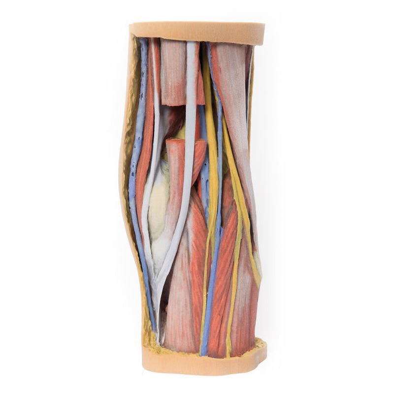
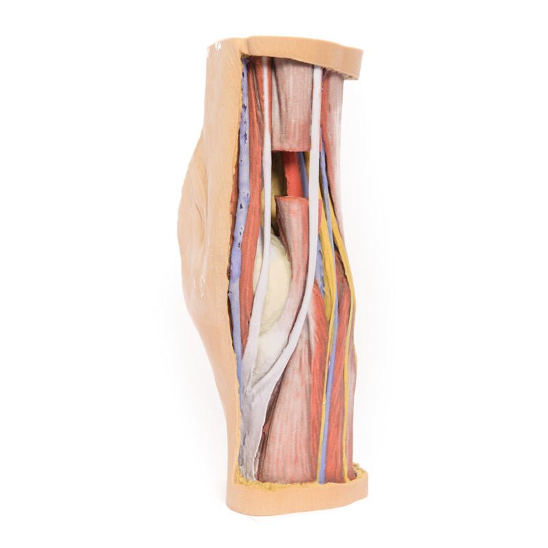
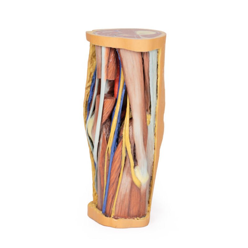
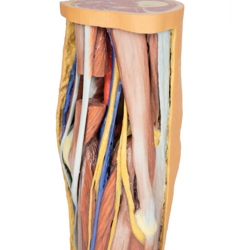
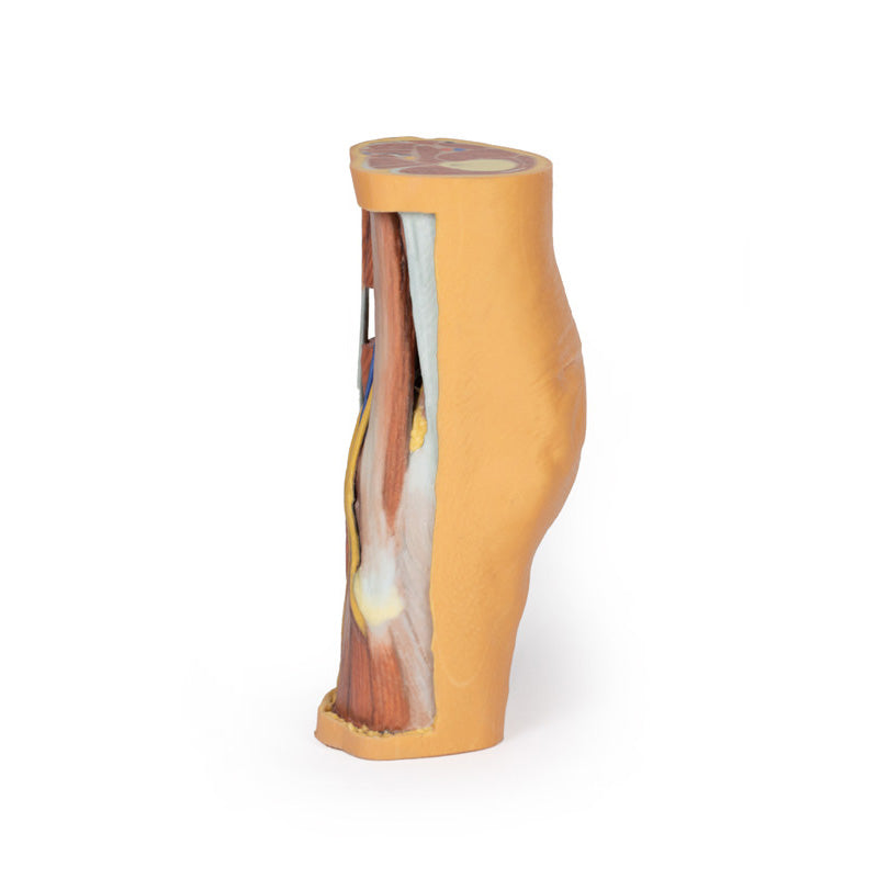
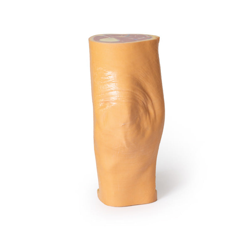
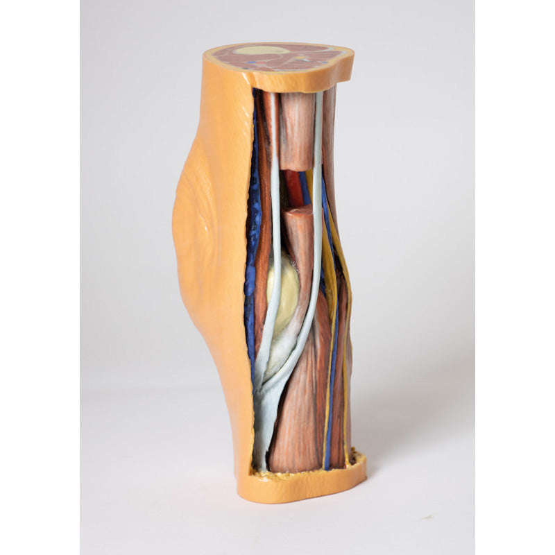
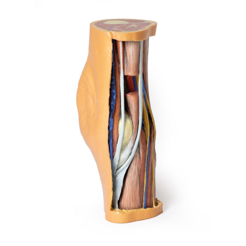
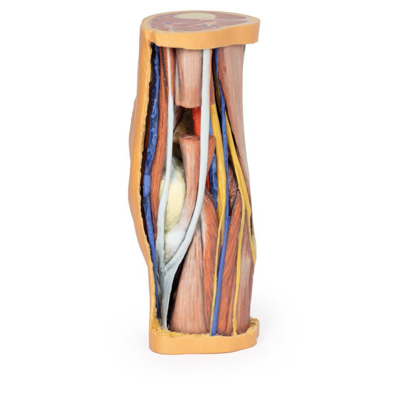
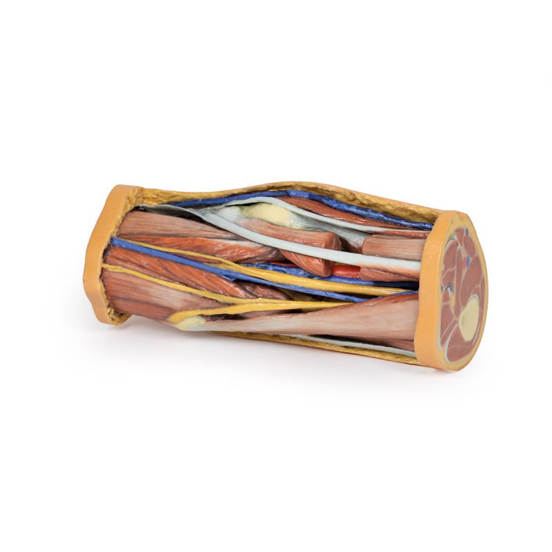
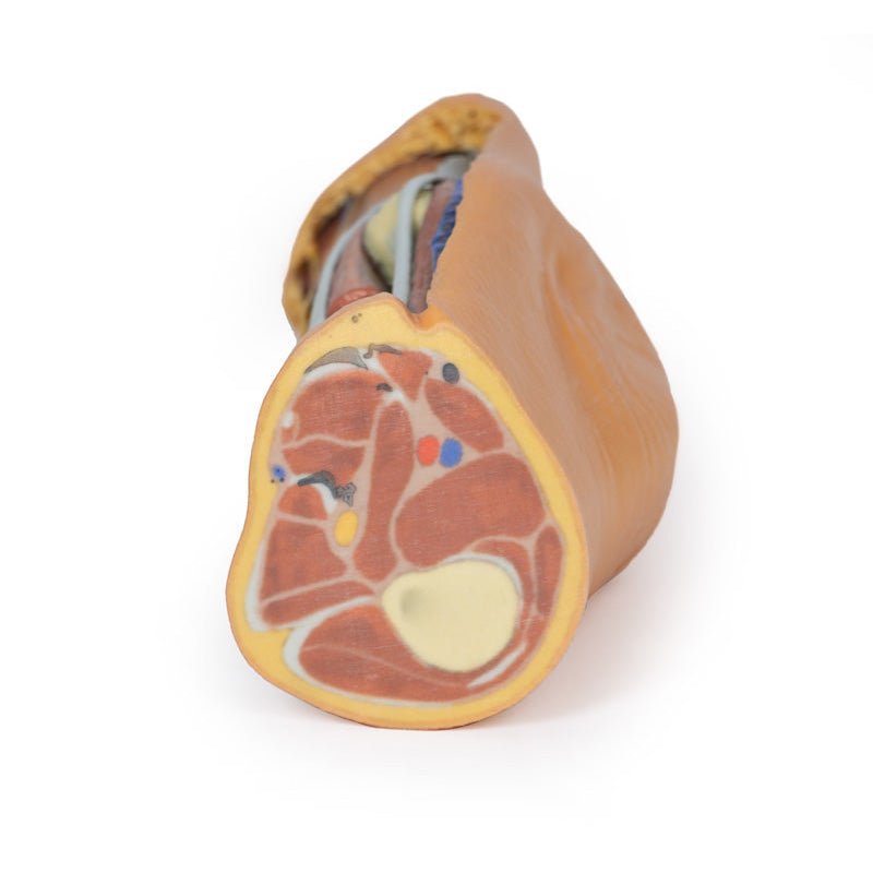
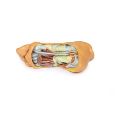
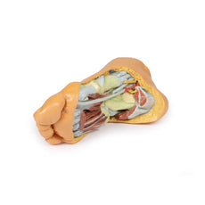
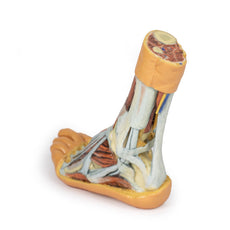
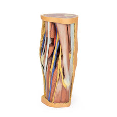
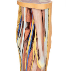
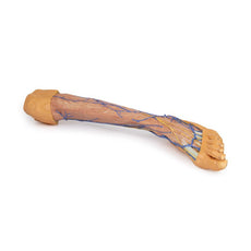
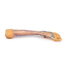
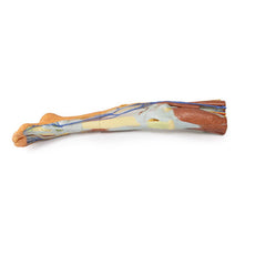
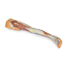
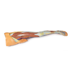
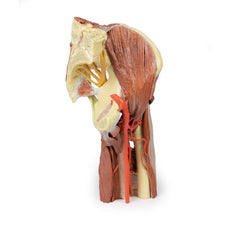
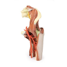
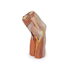
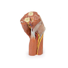
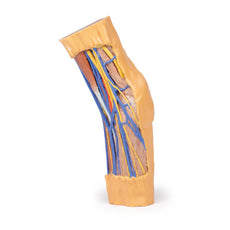
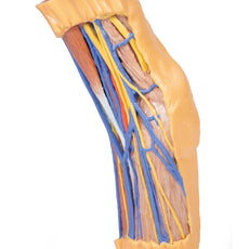
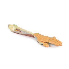
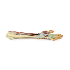
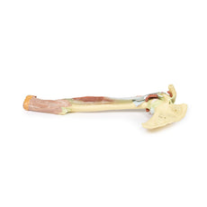
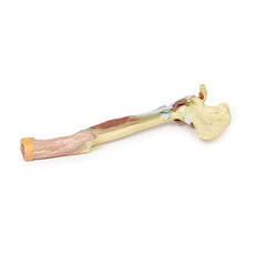
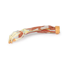
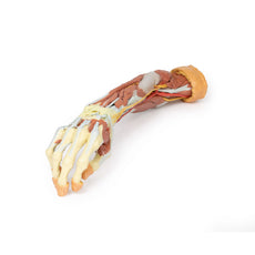
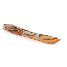
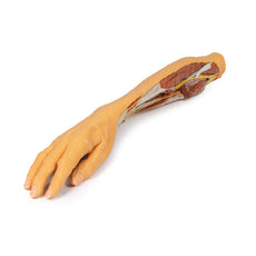
This 3D printed specimen preserves the distal thigh and proximal leg, dissected posteriorly to demonstrate the contents of the popliteal fossa and surrounding region. The proximal cross-section demonstrates the anterior, posterior and medial compartment muscles, with the femoral artery and vein visible within the adductor canal.The sciatic nerve and great saphenous vein are also visible. The skin, superficial fascia, and de
3D Printed Popliteal Fossa Model
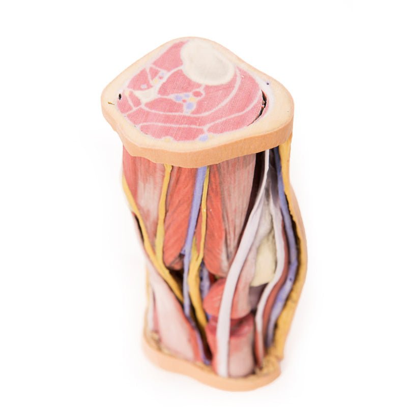
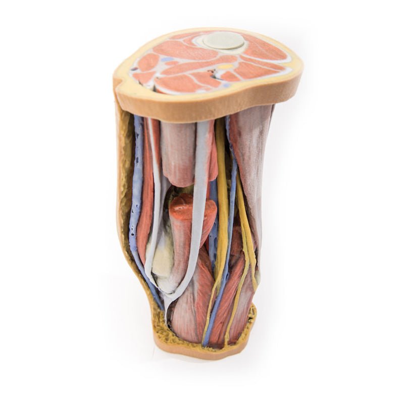
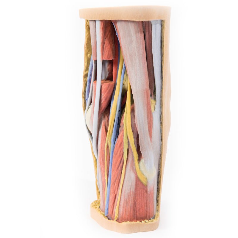
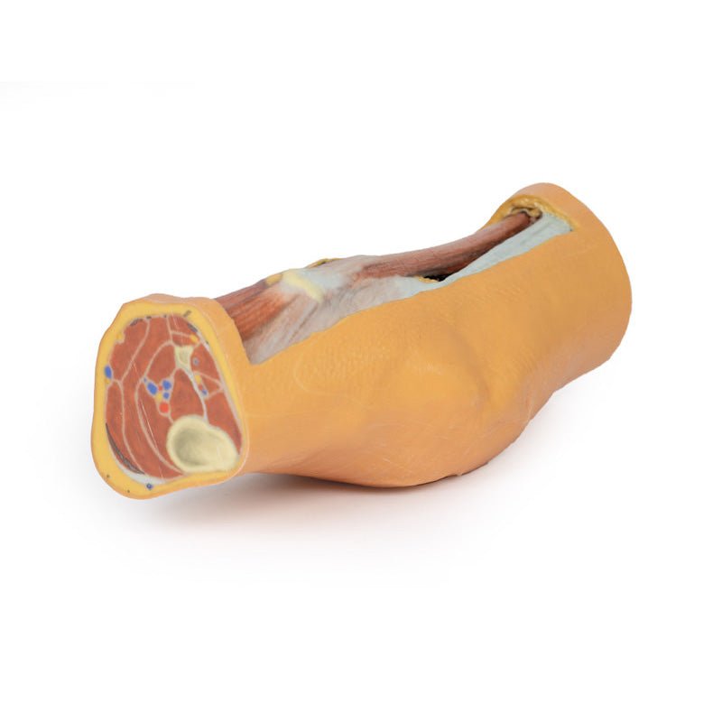
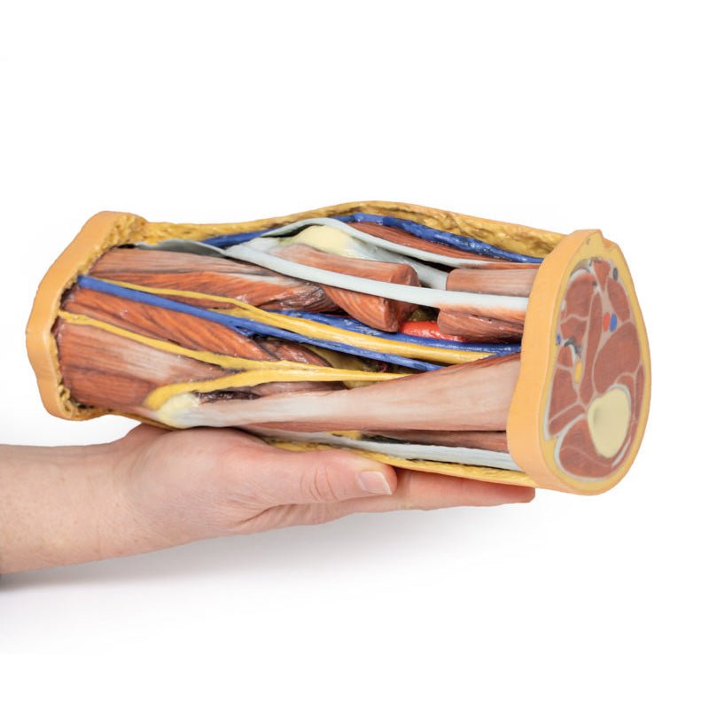
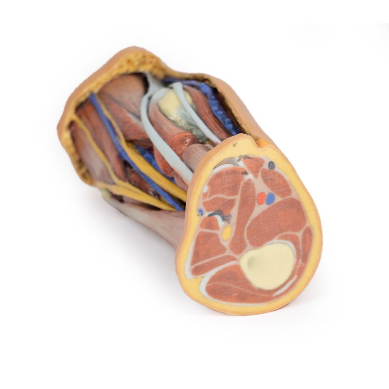
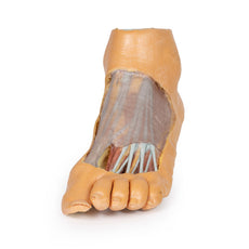
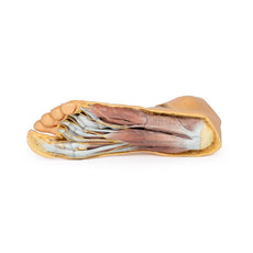
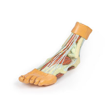
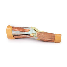
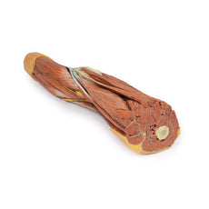
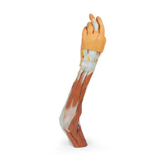
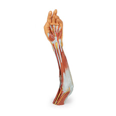
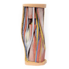
This 3D printed specimen preserves the distal thigh and proximal leg, dissected posteriorly to demonstrate the contents of the popliteal fossa and surrounding region. The proximal cross-section demonstrates the anterior, posterior and medial compartment muscles, with the femoral artery and vein visible within the adductor canal.The sciatic nerve and great saphenous vein are also visible. The skin, superficial fascia, and deep fascia have been removed over the popliteal fossa to expose the contents of the space. The muscular borders of the space are intact except for a window cut into the semimembranosus muscle to allow a view of the popliteal artery and vein near the adductor magnus. On the medial aspect of the window the great saphenous vein descends on the surface of the sartorius muscle. Distally the sartorius is visible joining the semitendinosus and semimembranosus muscles to form the pes anserinus.
All major deep and superficial nerves and vessels of the space are visible, including the superior lateral genicular artery passing towards the anterior compartment of the thigh. Along the lateral margin the posterior aspect of iliotibial tract is visible descending to the lateral epicondyle of the tibia. The distal cross-section demonstrates the continuation of popliteal contents and branches. The great saphenous and small saphenous veins are visible within the superficial fascia, as are the medial and lateral sural cutaneous nerves.
Between the muscles of the posterior, lateral, and anterior compartments are the neurovascular bundles of the leg (posterior tibial artery, veins and tibial nerve; peroneal artery and veins; anterior tibial artery, veins and deep peroneal nerve; superficial peroneal nerve).
Download Handling Guidelines for 3D Printed Models
GTSimulators by Global Technologies
Erler Zimmer Authorized Dealer
The models are very detailed and delicate. With normal production machines you cannot realize such details like shown in these models.
The printer used is a color-plastic printer. This is the most suitable printer for these models.
The plastic material is already the best and most suitable material for these prints. (The other option would be a kind of gypsum, but this is way more fragile. You even cannot get them out of the printer without breaking them).The huge advantage of the prints is that they are very realistic as the data is coming from real human specimen. Nothing is shaped or stylized.
The users have to handle these prints with utmost care. They are not made for touching or bending any thin nerves, arteries, vessels etc. The 3D printed models should sit on a table and just rotated at the table.


