Your shopping cart is empty.
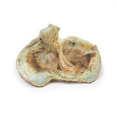
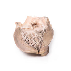
3D Printed Acute Bacterial Endocarditis
Item # MP2041Need an estimate?
Click Add To Quote

-
by
A trusted GT partner -
3D Printed Model
from a real specimen -
Gov't pricing
Available upon request
3D Printed Acute Bacterial Endocarditis
Clinical History
A 15-year old boy with cough and sputum developed a hectic (spiking) fever and chest pain a few
days before being admitted in a comatosed condition. Examination revealed an early diastolic murmur at the aortic
area, which radiated down the left sternal edge. He deteriorated very quickly and died, despite antibiotic
chemotherapy. Blood cultures grew Staphylococcus aureus.
Pathology
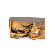
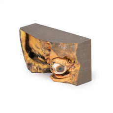
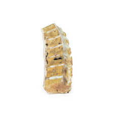
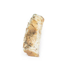
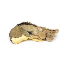
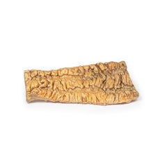
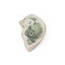
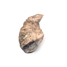
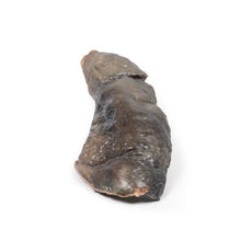
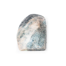
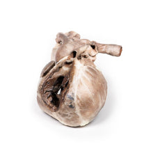
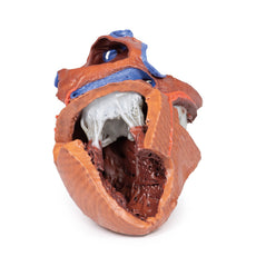
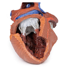
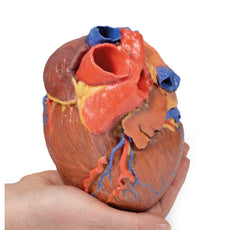
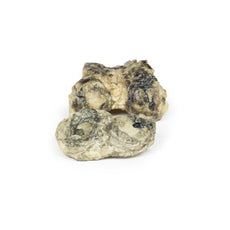
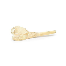
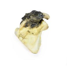
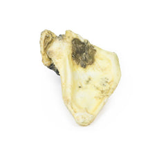
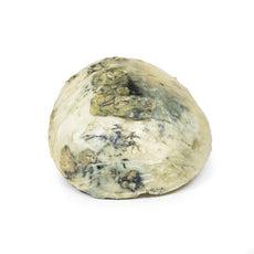
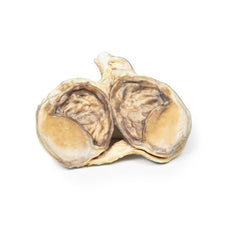
This small heart displays the left ventricle and associated valves. The non-coronary cusp of the aortic
valve is ulcerated and perforated and has friable vegetations attached. Immediately below this cusp a perforation
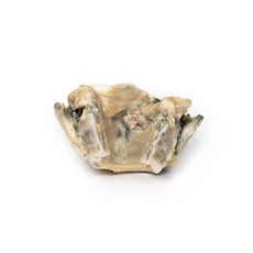
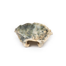
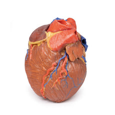
extends into the right atrium just above the tricuspid valve (see back of specimen. The other aortic cusp is also
thickened. This is an acute bacterial endocarditis with aortic cusp and atrioventricular perforations.
Further Information
Acute bacterial endocarditis is a form of infective endocarditis. Endocarditis due to fungal
infections can also occur although they are rare.
In normal circumstances, the endothelial lining of the heart
and valves is relatively resistant to infection by most bacteria or fungi. Therefore, in order for infective
endocarditis to occur, there needs to be initial damage or injury to the endocardial tissue. This often results in
aggregation of platelets and fibrin, which then become infected, resulting in vegetation formation (i.e. an
infective nidus). Staphylococcus aureus, however, is highly virulent and can sometimes infect normal heart valves.
After the initial platelet-fibrin aggregation, there is further activation of the coagulating system via the
extrinsic clotting pathway and initiation of the inflammatory response via monocytes, resulting in further growth of
the vegetation/thrombus. The microbial growth tends to occur within the fibrin matrix, which makes it difficult for
the immune responses to eradicate the infection. An additional problem is that these infected thombi can also
embolise causing distant sites of infection in smaller capillaries (e.g. in the kidney).
Risk factors for
developing an infective endocarditis include valvular heart disease, such as previous rheumatic heart disease,
congenital heart disease (e.g. ventricular septal defect or bicuspid aortic valve), prosthetic heart valves or any
previous invasive cardiac procedures. Anti-thrombotic medications – e.g. heparin or aspirin – may have to be
administered in patients at risk. Diagnosis is initially made by clinical examination followed by pathology (blood
cultures) and diagnostic imaging. Transthoracic echocardiogram is often first line, followed by transoesophageal
echocardiogram. Treatment includes antimicrobial therapy, anti-coagulants, and in some complicated cases, surgical
intervention such as valve surgery.
 Handling Guidelines for 3D Printed Models
GTSimulators by Global Technologies
Erler Zimmer Authorized Dealer
The models are very detailed and delicate. With normal production machines you cannot realize such details like shown in these models.
The printer used is a color-plastic printer. This is the most suitable printer for these models.
The plastic material is already the best and most suitable material for these prints. (The other option would be a kind of gypsum, but this is way more fragile. You even cannot get them out of the printer without breaking them).The huge advantage of the prints is that they are very realistic as the data is coming from real human specimen. Nothing is shaped or stylized.
The users have to handle these prints with utmost care. They are not made for touching or bending any thin nerves, arteries, vessels etc. The 3D printed models should sit on a table and just rotated at the table.


