Your shopping cart is empty.
Female Dog Phantom for X-Ray and Ultrasound Imaging
Item # TPS-DG-A01Need an estimate?
Click Add To Quote

-
by
A trusted GT partner -
FREE Shipping
U.S. Contiguous States Only -
2-Year Warranty
Provided by manufacturer -
Gov't pricing
Available upon request -
Download Manual
Click to view product's manual
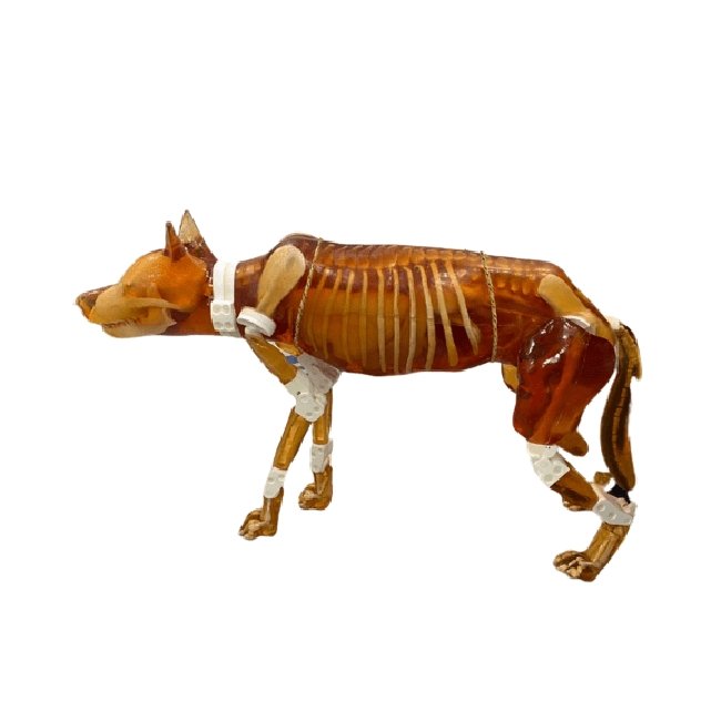
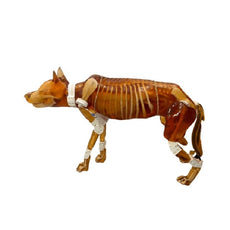
Female Dog Phantom for X-Ray and Ultrasound Imaging
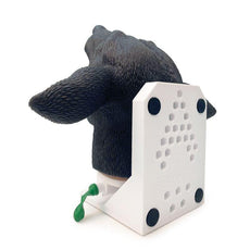
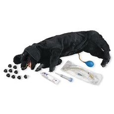
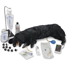
The newly designed detachable dog phantom serves as an independent training simulator compatible with ultrasound and
X-Ray/CT imaging. It is an ideal teaching tool for veterinary professionals, featuring improved anatomical
structures and removable body parts. This allows for practicing various positioning techniques under both imaging
modalities.
The newly designed detacha
Female Dog Phantom for X-Ray and Ultrasound Imaging
The newly designed detachable dog phantom serves as an independent training simulator compatible with ultrasound and
X-Ray/CT imaging. It is an ideal teaching tool for veterinary professionals, featuring improved anatomical
structures and removable body parts. This allows for practicing various positioning techniques under both imaging
modalities.
The newly designed detachable dog phantom is a training simulator independent of external hardware/software. The
phantom is compatible with Ultrasound and X-RAY/CT imaging. It has all the right features to be an ideal teaching
tool for sonographers, radiographers, veterinary residents, and other vet professionals.
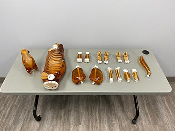
Unlike its predecessor, the new detachable dog phantom has improved anatomical structures and removable body parts
(head, limbs, torso, and tail). This improvement is an added advantage as it helps perform various positioning
techniques under the US and X-RAY/CT imaging modalities.
ANATOMY
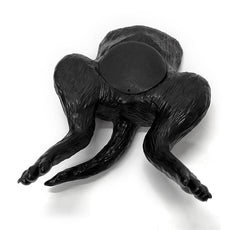
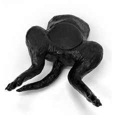
Adult Dog Front Arms
- Arm (Humerus)
- Elbow Joints
- Forearm (Radius, Ulna)
- Paw (Wrist with Fingers)
- Skin-Mimicking Material
Adult Dog Torso
- Complete Spine
- Complete Ribcage
- Shoulder (Scapula Bones)
- Pelvis
- Heart
- Blood Vessels Connected to Heart
- Lungs
- Diaphragm
- Stomach
- Liver and Gallbladder
- Kidneys
- Spleen
- Pancreas
- Arteries for Kidneys
- Bladder
- Ureters and Uterine
- Large and Small Intestine
- Dog Phantom
- User Manual/Assembly Instructions
- 2-Year Warranty & Unlimited Customer Service
- Hard Carry Case

- Height: 33 x 18 x 6 in (approx.)
- Weight: 40 lbs (approx.)
- Height: 32 x 23 x 13 in (approx.)
- Weight: 99 lbs (approx.)
- Soft tissue and organs: Composition of urethane-based soft resin
- Synthetic bones: Patented ceramic-reinforced epoxy-based composite material
GTSimulators by Global Technologies
True Phantom Solutions Authorized Dealer
Female Dog Phantom for X-Ray and Ultrasound Imaging
Phantom:- Height: 33 x 18 x 6 in (approx.)
- Weight: 40 lbs (approx.)
- Height: 32 x 23 x 13 in (approx.)
- Weight: 99 lbs (approx.)
- Soft tissue and organs: Composition of urethane-based soft resin
- Synthetic bones: Patented ceramic-reinforced epoxy-based composite material
| Type of Tissue | Sound Velocity[m/s] | Density [g/cm3] | Attenuation Measured at 2.25 MHz [dB/cm] | Hardness [Shore OO] | T2 [ms] | Speckles |
|---|---|---|---|---|---|---|
| Organs with Speckles (liver, kidneys, etc.) | 1400 ± 10 | 0.99 | 1.0 ± 0.2 | 20 | 70 | VARIABLE |
| Organs without Speckles (stomach, intestines, etc.) | 1400 ± 10 | 0.99 | 1.0 ± 0.2 | 20 | 70 | NO |
| Body Tissue | 1400 ± 10 | 1.00 | 1.2 ± 0.2 | 30 | 65 | NO/LOW |
| Cortical Bone | 3000 ± 30 | 2.31 | 6.4 ± 0.3 | N/A | N/A | N/A |
| Trabecular Bone | 2800 ± 50 | 2.03 | 21 ± 2 | N/A | N/A | N/A |
| Brain Matter | 1400 ± 10 | 0.99 | 1.0 ± 0.2 | 20 | 70 | YES |
| Thermal Conductivity | Volumetric Specific Heat Capacity | Thermal Diffusivity | Thermal Resistivity | Specific Heat | Speed of Sound |
|---|---|---|---|---|---|
| 0.776 W/ m K | 1.040 MJ/ m^3 K | 0.746 mm^2/ s | 1.289 m K/ W | 0.978 J/ g Deg Celsius |
|
| S.No. | Tissue Type | HU Value (Average) |
|---|---|---|
| 1 | Body Tissue | -25 |
| 2 | Brain Tissue | -25 |
| 3 | Trabecular Bone | 800 |
| 4 | Cortical Bone | 1300 |
| 5 | Aorta | 40 |
| 6 | Vena Cava | 40 |
| 7 | Trachea | 80 (Tissue Part),-1000 (Air Filled Part) |
| 8 | Pancreas | 110 |
| 9 | Spleen | 110 |
| 10 | Kidneys | 110 |
| 11 | Bladder | 35 |
| 12 | Rectum Wall | 100 |
| 13 | Sigmoid Colon Wall | 100 |
| 14 | Heart | 40 |
| 15 | Liver | 110 |
| 16 | Gallbladder | 35 |
GTSimulators by Global Technologies
True Phantom Solutions Authorized Dealer


